Detectors capture emitted photons and convert them into electronic signals, making them one of the most influential
components for microscope performance and image quality.
Different detector technologies and models offer different strengths in terms of sensitivity, field of view, imaging
speed and compatibility with specific imaging modalities. Understanding detector specifications is therefore essential
when assessing whether a microscope configuration meets the requirements of your application.
This overview introduces the detector technologies commonly used in benchtop fluorescence microscopy and explains the
key specifications that influence imaging performance.
Which Detectors Are Used in Benchtop Fluorescence Microscopes?
Benchtop systems primarily use integrated sCMOS cameras (here: monochrome cameras) for widefield and spinning-disk confocal imaging.
In modular platforms, cameras can be exchanged and upgraded over time as applications evolve or as new sensor
technologies become available.
Detectors of benchtop systems connected to point-scanning confocal microscopes employ photomultiplier tubes (PMTs),
including GaAsP variants.
| sCMOS cameras |
PMTs (Photomultiplier tubes) |
- High quantum efficiency (typically 70–95%), enabling sensitive detection of weak signals
- Large
sensor formats support large fields of view and reduce the number of tiles needed per dataset
- Low read noise
and low dark current support fast, low-noise acquisition
- Pixel sizes around 6.5 µm provide efficient sampling
under Nyquist conditions while supporting fast imaging
- Used in widefield and spinning-disk confocal
microscopes
- Suitable for nearly all fluorescence imaging applications
|
- Quantum efficiency up to ~45% for GaAsP PMTs
- Intrinsic dark current values around 2e-
- Detect
light from one pixel at a time during laser scanning
- Used in point-scanning confocal microscopes
|
Which Detector Specifications Should I Consider in a Benchtop Fluorescence Microscope?
| Key detector parameters |
Description |
| Quantum Efficiency (QE) |
- Percentage of incoming photons captured and converted into photoelectrons (e.g. 50% QE = 50% of
photons are converted to photoelectrons)
- Wavelength-dependent, with most sensors optimised for the visible spectrum
- Higher QE corresponds to higher sensor sensitivity
- QE values above 70% are recommended for most fluorescence applications
|
| Pixel size |
- Physical dimensions of a single sensor pixel (light-sensitive element) in µm
- Must satisfy Nyquist sampling criteria to capture fine structural detail
- Trade-off between resolution and signal intensity, especially relevant at lower magnifications:
- Smaller pixels = higher spatial resolution, but less light collected per pixel
- Larger pixels = higher signal per pixel but lower spatial resolution
- Pixel binning may be used to increase effective pixel size and improve signal-to-noise ratio at
expense of image sampling
|
| Sensor size |
- Determines the maximum field of view that can be captured in a single image frame
- Large sensors capture more data in one image, reducing the number of tiles needed for large
samples (organoids, tissues, multi-well plates)
- Smaller sensors effectively "crop" the microscope's image circle, which can
increase acquisition speed but reduces imaging efficiency
- The usable sensor area may be limited by the microscope's field of view, determined by the
objective, internal optics and camera adapter
|
| Read noise |
- Noise within the camera, introduced during analogue-to-digital conversion and signal
amplification processes
- Lower read noise equals a lower noise floor and improved detection of weak fluorescence signals
|
| Dark current |
- Signal generated internally within the sensor in the absence of light (e-/pixel/second)
- Becomes more important at extended exposure durations (e.g. luminescence) and low-signal
experiments
- Camera cooling (active or passive) helps reduce dark current, so it does not impact image quality
during routine imaging
|
| Cooling |
- Reduces dark current and hot (bright) pixels and improves signal-to-noise ratio
- Implemented as passive cooling (heat dissipation from camera body to ambient air), or active
cooling (fan-based or liquid cooling)
- Air cooling might induce small vibrations but is an effective solution for many benchtop
applications when controlled properly
- Liquid cooling achieves deeper cooling for the lowest dark current, but is typically not used in
benchtop systems
|
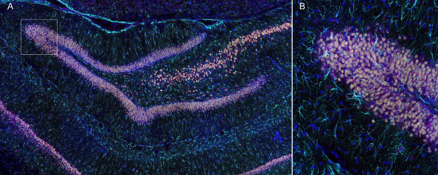
Fig. 1 – Large-scale imaging of rat hippocampus. Using BC43 confocal imaging combined with a
high-quantum-efficiency (QE) sCMOS detector, a large rat hippocampal tissue sample was rapidly acquired, capturing a
total of 2,240 images in just 12 minutes. (A) A 4-channel composite image consisting of 28 tiled fields across a
20-plane Z-stack, providing a comprehensive volumetric view of the rat hippocampus. (B) A high-resolution zoomed
region highlighting fine neural structures and cellular detail. The tissue section was stained with markers
indicative of oxidative stress. Sample courtesy of Dr. Daniela Ostrowski, Department of Biology, Truman State
University.
Exploring Benchtop Fluorescence Microscopes?
Learn more about the key technologies, components and practical considerations involved in selecting a benchtop
fluorescence microscope.
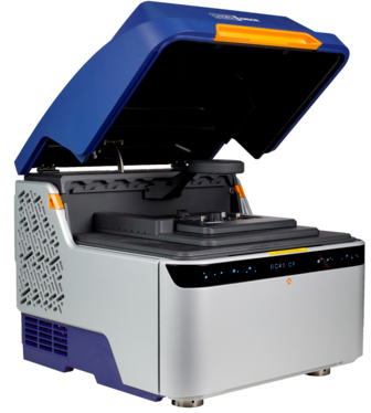
BC43 – Exceptional Performance, Certified Quality, High Productivity
The BC43
features a high sensitivity sCMOS camera designed for high-quality imaging across a wide range of applications.
• High QE (82%) and very low read noise for high sensitivity
• High frame rates for capturing dynamic processes
• Wide field of view for efficient large-area imaging
• Active air cooling to minimise dark current and hot pixels