The illumination source plays a central role in fluorescence microscopy, as it determines compatibility with
different imaging modes, spectral flexibility, image contrast, and resolution. In fluorescence microscopes, excitation
stability and illumination uniformity are particularly important, as poor performance in these areas directly
compromises image quality. This overview compares LED and laser options in benchtop fluorescence microscopes.
Choosing a Benchtop Fluorescence Microscope – Which Illumination Source Should I Consider?
Benchtop fluorescence microscopes may integrate LEDs, lasers, or a combination of both. The choice of light source
influences not only imaging performance, but also system cost and maintenance requirements.
LEDs
- Preferred illumination source for widefield fluorescence
- Have largely replaced historically used Xenon and metal-halide lamps*
- Offer long lifetimes with precise and stable output over extended times
- Require minimal maintenance
- Available across multiple excitation wavelengths, supporting multi-colour imaging
- Deliver gentle illumination that helps reduce photobleaching, making them well-suited for live-cell imaging
- Produce very little heat, minimising thermal drift, an advantage especially in compact, enclosed benchtop systems
- Limitations:
- Low efficiency in the ultraviolet (UV) range
- Higher initial costs, especially when many wavelengths are required (typically one LED per colour)
* Xenon and metal halide lamps are not discussed further in this overview, as they are generally not utilised in
modern systems and not relevant for modern benchtop imaging systems.
Lasers
- Essential illumination source for confocal microscopy applications
- High power and well-defined beam characteristics:
- Monochromatic: very narrow wavelength range
- Directional: beam propagates along a straight line in a defined direction
- Coherent: Fixed and predictable phase relationship in space and time
- Provide beam properties required for focusing through pinholes, enabling high-quality optical sectioning
- Available as single-mode or multi-mode lasers:
- Single-mode lasers: Tightly focused beams, ideal for point-scanning confocal microscopes
- Multi-mode lasers: available generally in higher power modules. Offered in a variety of
wavelengths and power levels
- The combination of installed lasers defines the available fluorescence channels (together with the emission
filters and dichroic mirror configuration)
- Can be used for both widefield and confocal illumination, reducing system complexity and long-term maintenance in
multimodal benchtop systems
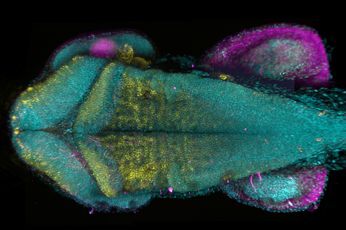
Fig. 1 – Image deep into the zebrafish brain with BC43. The combination of Oxford Instruments' patented
Borealis illumination technology, dual microlens optics, and a highly accurate XZ stage enables acquisition of
multiple XYZ planes throughout the zebrafish brain, which are seamlessly stitched into a single, continuous image.
The image shows a maximum projection of zebrafish hindbrain at 48hpf staining the neurons (anti-HuC/D serum,
yellow), glia (anti-zrf serum, magenta). 30 tiles were acquired to compose the image; each tile had 175 slices, over
a 370 mm Z range. Image credits: Marco Campinho, Universidade do Algarve.
Exploring Benchtop Fluorescence Microscopes?
Learn more about the key technologies, components and practical considerations involved in selecting a benchtop
fluorescence microscope.
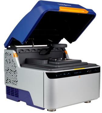
BC43 – Exceptional Performance, Certified Quality, High Productivity
BC43 uses
multimodal lasers integrated through the patented Borealis illumination system, ensuring uniform illumination across
the field of view during imaging in the confocal mode.
← Back to Learning