The microscope chassis (body) defines the system's overall architecture and strongly influences sample
compatibility, environmental stability, illumination setup, and workflow integration. While it does not usually
dominate the total system cost, it determines many practical aspects of usability, flexibility, and overall imaging
performance.
This overview explains the microscope architectures and design choices found in benchtop systems and highlights the
key factors to consider when evaluating a microscope.
Upright vs Inverted Microscopes
| Configuration |
Description |
| Upright microscopes |
- Compact layout with objectives positioned above the sample stage.
- Preferred for larger specimens
and fixed samples.
- Typically simpler in design than inverted microscopes.
- Well-suited for teaching purposes.
|
| Inverted microscopes |
- Objectives positioned below the microscope stage.
- Ideal for live-cell imaging and samples in
aqueous media (cells, organoids, microtissues).
- Preferred configuration for integration with micromanipulation
setups.
|
Open-framed vs Enclosed/Boxed Designs
| Configuration |
Description |
| Open-framed microscopes |
- Provide direct physical access to the sample and optical components.
- More susceptible to
environmental influences such as ambient light, temperature drift, and vibrations.
- Generally easier to maintain
and upgrade.
|
| Enclosed/boxed microscopes |
- Shield optical components and samples from ambient light.
- Offer partial protection from
environmental fluctuations, dust and optical contaminants.
- Enable safer laser handling and protect system
optics.
- Particularly suitable for less-experienced users and multi-user environments.
|
Eyepiece vs Digital-only Systems
| Configuration |
Description |
| Eyepiece-based systems |
- Enable direct sample inspection and manual navigation.
- Familiar and intuitive sample
visualisation for many users.
- More challenging to share observations with a group.
|
| Digital-only systems |
- More ergonomic and comfortable for extended use.
- Provide a larger effective viewing area via
screen display.
- Simplify documentation and collaboration (images are immediately available digitally).
- Streamline automated scanning and workflow needs.
- Not all systems support high-magnification imaging.
|
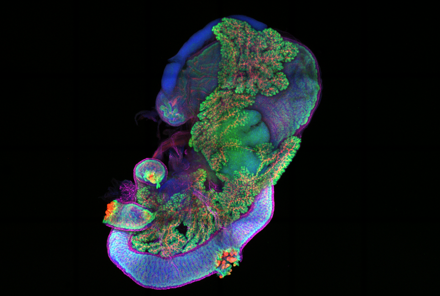
Fig. 1 — Mouse pancreas and connected tissues imaged with BC43. Whole-mount immunofluorescence staining of a
whole E16 mouse pancreas, with the spleen, stomach and duodenum intact, enabling further investigation of the
anatomical characteristics in pancreatic development and disease. The tissue was imaged using a confocal microscope
by tiling and processed using Imaris. Image courtesy of Cedric Yu, PhD, University of California, San
Francisco.
Exploring Benchtop Fluorescence Microscopes
Learn more about the key technologies, components and practical considerations involved in selecting a benchtop
fluorescence microscope.
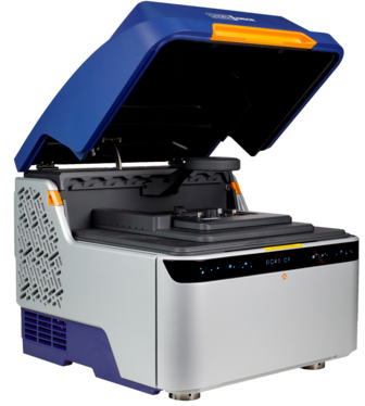
BC43 – Exceptional Performance, Certified Quality, High Productivity
BC43 is a
fully digital, enclosed microscope with an inverted beam path. It enables intuitive sample navigation using a 2x
objective, while the integrated high-QE sCMOS camera supports sensitive and fast imaging across a wide magnification
range, including high-magnification imaging up to 100x