Fluctuating environmental conditions and vibrations can have a significant impact on image quality, reproducibility
and experimental success in fluorescence microscopy. Especially long-term studies, live-cell experiments,
high-resolution imaging, and automated imaging workflows require mechanical stability and careful control of
environmental conditions such as temperature and humidity.
Different microscope designs offer varying levels of vibration reduction and environmental regulation. This overview
explains the most common solutions on vibration and environmental control and highlights key considerations when
selecting a system.
Vibrational Control
Stable mechanical conditions ensure the acquisition of sharp, reproducible images, particularly during long
time-lapse, Z-stack, 3D, or high-resolution imaging experiments. Depending on the imaging modality used and the
vibration profile of your laboratory environment, vibration-minimising features can be critical.
While widefield fluorescence imaging, especially at lower magnifications, often does not require dedicated vibration
control, confocal and super-resolution imaging are more sensitive to mechanical disturbances and need
vibration-reducing measures.
| Configuration |
Description |
| Passive vibration control |
- Relies on rigid system frames, damping materials, or pneumatic isolators to absorb or decouple
bench-borne vibrations.
- Provides sufficient mechanical stability for most benchtop applications including
high-resolution widefield, confocal, and super-resolution imaging with resolutions down to ~100 nm.
|
| Active vibration control |
- Uses sensors and actuators to detect and counteract vibrations in real time.
- Extremely relevant
for single-molecule localisation microscopy (SMLM).
- Introduces higher system complexity and costs compared to
passive solutions.
|
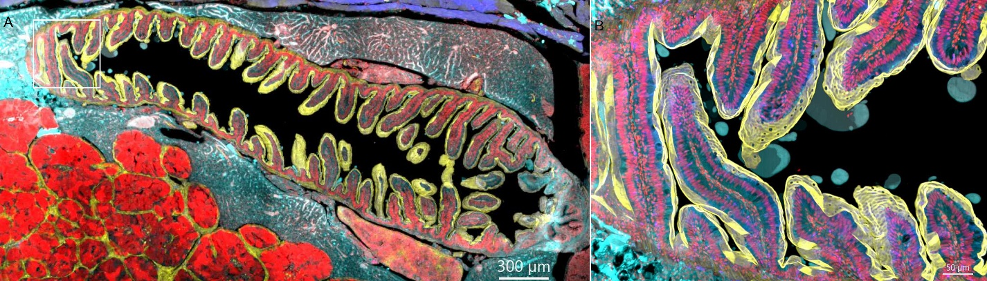
Fig. 1 — Stitched image of zebrafish intestine. The image was acquired using the confocal imaging modality of the
BC43 system, employing 4 imaging channels, 77 z-stacks, and 28 tiles. The final stitched image comprises a total of
15,092 individual images, yet appears as a single seamless snapshot. This high-quality stitching is achieved through
Borealis's uniform illumination, robust anti-vibration design, and the Imaris Stitch algorithm. (A) Full view of
the intestine section. (B) Zoomed-in view highlighting structural details of the intestinal tissue. Both
deconvolution and stitching options were enabled during acquisition. Sample courtesy of Julien Resseguier, NorMic,
University of Oslo. Image credit: Claudia Florindo, Oxford Instruments.
Environmental Control
Maintaining stable environmental conditions is essential for reliable and reproducible imaging, particularly when
working with live cells or long time-lapse experiments. Fluctuations in temperature, gas composition, humidity, or
mechanical stability can introduce image drift and directly affect cell physiology.
Benchtop fluorescence microscopes differ considerably in the level of environmental control they provide. Enclosed
(boxed) systems naturally reduce exposure to ambient environmental variations compared with open-frame designs.
However, for specific applications such as live-cell and long-term imaging, environmental control solutions are
required.
| Configuration |
Description |
| Integrated environmental control |
- Built directly into the microscope system.
- Provide stable regulation of temperature, gas
composition (CO2/O2), and humidity.
- Eliminate the need for additional devices,
simplifying system setup.
- Increase the initial cost of the overall imaging system.
- Usually not compatible
with strict decontamination procedures required in BSL-3/4 environments.
|
| Stage-top incubators |
- External incubation chambers mounted directly on the frame of the microscope stage.
- Can be
added or removed as required.
- Provide temperature, humidity, and gas control and often support perfusion.
- Offer high flexibility, making them well suited for multi-user labs and diverse workflows.
- Can be available
with BSL-3/4-compatible decontamination options (availability depends on manufacturer and must be specified).
|
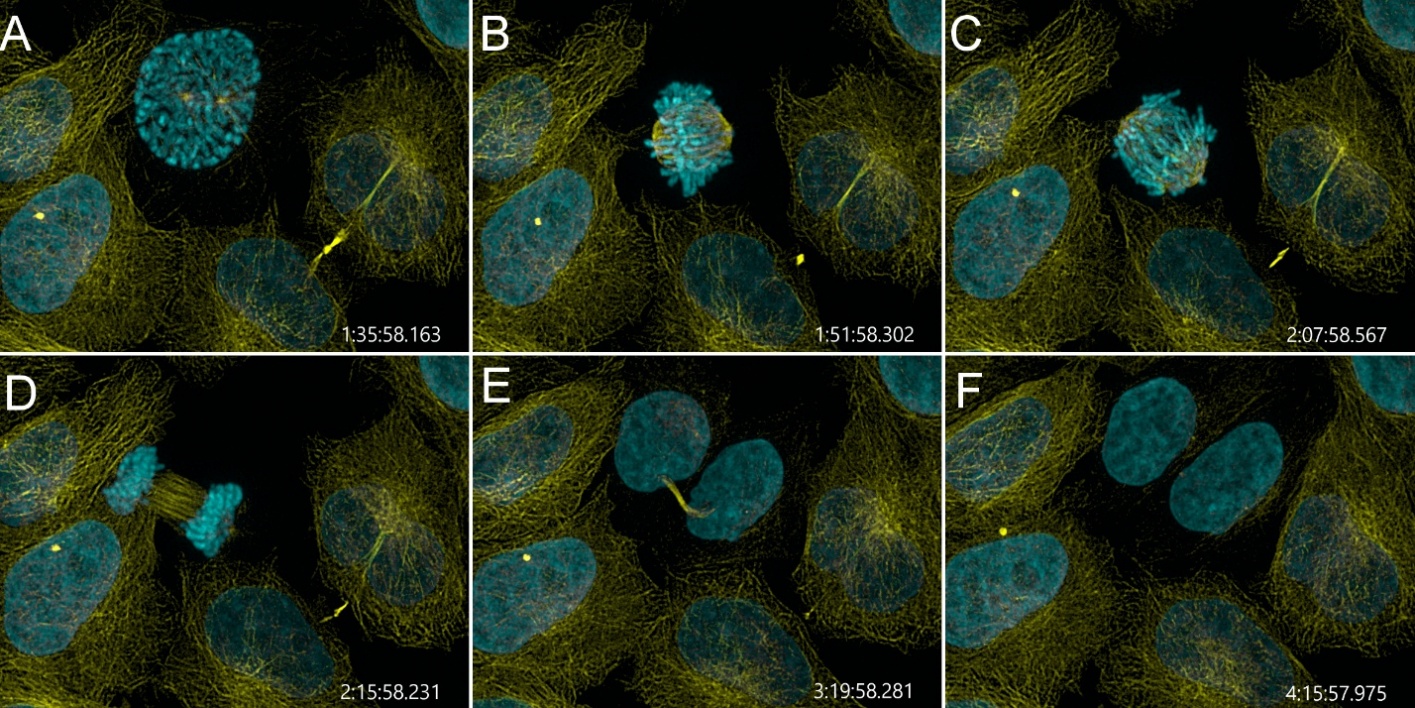
Fig. 2 — Visualising mammalian cell division with BC43. Time lapse image acquired every 3 min, using the confocal
imaging modality. Cells were incubated at 37°C, Yellow: microtubules; cyan: DNA. Image credit: Ines Baião-Santos and
Álvaro Tavares, Universidade do Algarve, Claudia Florindo, Oxford Instruments.
Exploring Benchtop Fluorescence Microscopes?
Learn more about the key technologies, components and practical considerations involved in selecting a benchtop
fluorescence microscope.
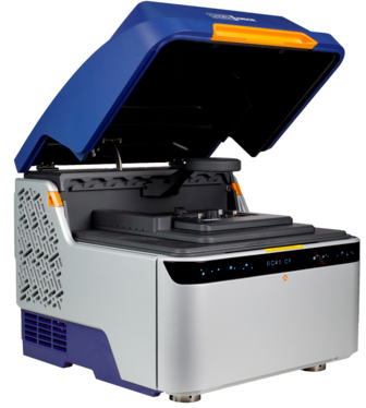
BC43 – Exceptional Performance, Certified Quality, High Productivity
- The BC43 is mounted on feet composed of silicone dampers that provide passive vibration control, creating an
exceptionally robust anti-vibration setup for the entire system. When placed on a standard laboratory bench, routine
activities such as operating benchtop centrifuges or sample mixers can continue without compromising image
acquisition performance. High-quality 3D stacks, multi-tile stitched images and even super-resolution images can all
be acquired on the BC43.
- The BC43 supports stage-top incubators that integrate temperature, humidity, CO2, and hypoxia control,
allowing full experimental flexibility. For BSL-3/4 laboratories, the BC43 incubation solution specifically includes
a coated stage-top incubator that can withstand robust decontamination protocols (decontamination procedures should
be confirmed in advance).