Choosing between a colour and a monochrome camera for microscopy is one of the most important decisions in scientific imaging, and one that needs made before we consider different sensor formats and other specific parameters. Both camera types may use similar CMOS or sCMOS sensor architectures, but the way they capture signals is different. This significantly affects sensitivity, spatial resolution, colour fidelity, quantitative accuracy and workflow. Here we provide a brief overview of both colour and monochrome cameras, and why we typically use monochrome cameras for fluorescence microscopy applications.
How Colour Cameras Capture Images
Most colour cameras used in consumer and scientific imaging use a Bayer filter array placed over the sensor. In this configuration, a filter above each pixel will allow predominantly red, green or blue light to pass to the pixel below.
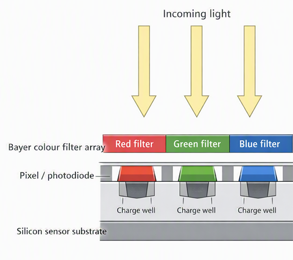
Figure 1: Representative image of a typical colour sensor with Bayer filter layer above pixels creating pixels sensitive to Red, Blue or Green.
Typically, the Bayer filtering is a repeating pattern with two green pixels for every red and blue pixel to mimic the response of the human eye which is more responsive to green. Image Signal Processing then reconstructs a full-colour image by interpolating the missing colour information at each pixel (demosaicing).
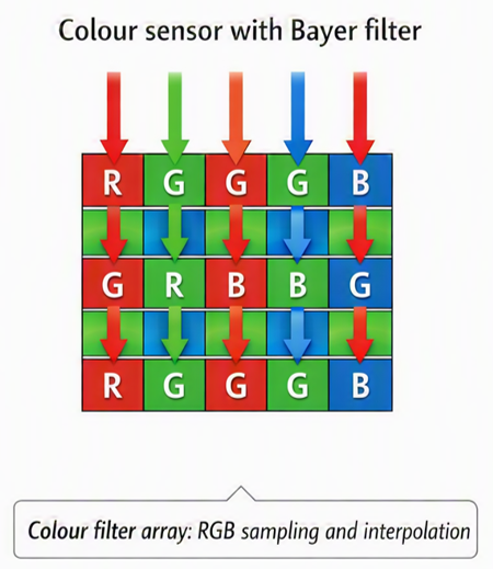
Figure 2: Representative image of a typical colour sensor with Bayer arrangement of Red (R), Blue (B), and Green (G) responsive pixels.
This approach provides RGB images that are easy to interpret, making colour cameras well suited to routine brightfield microscopy, histology, pathology, stained biological sections, materials inspection and education purposes.
There are true colour cameras available that do not use the Bayer pattern and have equal proportions of Red, Blue and Green sensitive pixels. These are used in pathology and materials inspection applications where true colour reproduction is essential.
The trade-off in colour cameras is that the colour filter array blocks much of the incoming light from each pixel. A red-filtered pixel, for example, captures red photons but rejects most green or blue photons that arrive at that pixel. This means a significant fraction of the available light does not hit a pixel that can detect it and thus will not contribute to the recorded signal. This can cut the detection performance by as much as 66-75%. As a result, colour cameras deliver notably lower sensitivity and weaker detection performance than monochrome cameras.
The process of demosaicing can also reduce effective spatial resolution. As fine detail is interpolated and reconstructed from neighbouring pixels rather than measured directly at every pixel, there are effectively gaps due to the wavelength filtering of the Bayer or other RGB pattern.
For these reasons, colour cameras are generally not suited to the low signal levels of typical fluorescence microscopy, where light levels and exposure times are intentionally kept as low as possible to minimise photobleaching and phototoxicity.
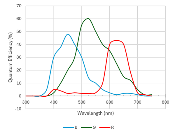
Figure 3: Representative spectral response curves for the red, green and blue filtered pixels in a colour sensor.
How Monochrome Cameras Capture Images
In contrast to colour cameras, monochrome cameras record intensity only, without inherent colour information. Differentiation between wavelengths therefore relies on optical filtering within the light path to select the signal before it reaches the detector.
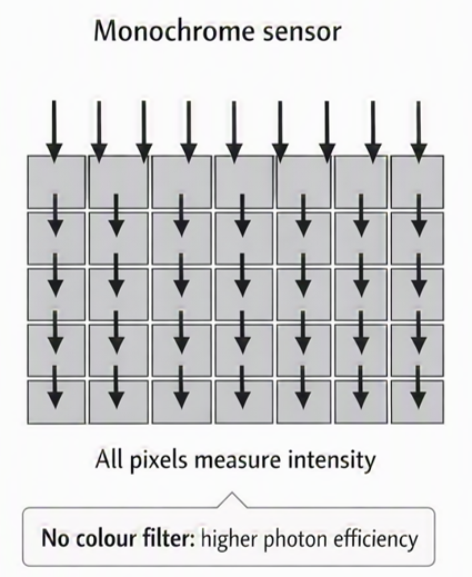
Figure 4: All pixels of the monochrome sensor capture the incoming signals with a high efficiency in the visible range.
Each pixel of a monochrome camera records light intensity across the sensor's full spectral response, so more photons can reach the photosensitive area. This gives monochrome cameras a significant sensitivity advantage in low-light applications such as fluorescence microscopy, live-cell imaging, calcium imaging, high-speed imaging and weak-signal detection. Furthermore, since every pixel in the sensor array is responsive to light it allows a much higher pixel density and allows more detail in the image to be maintained without any interpolation.
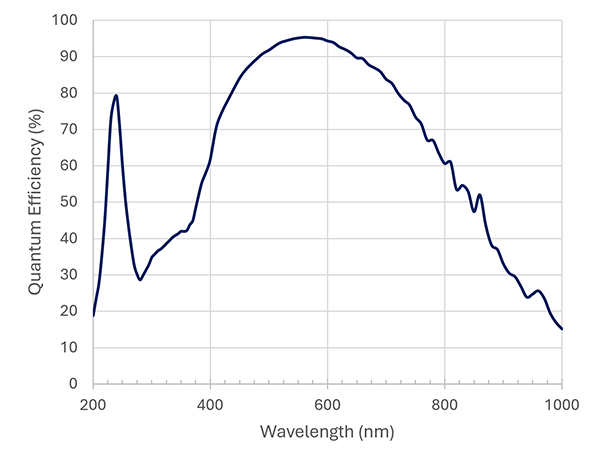
Figure 5: Back-illuminated monochrome cameras deliver the highest quantum efficiency (QE), with models such as the Sona sCMOS delivering peak QE of 95%.
Scientific grade (sCMOS) cameras feature more advanced electronics and supporting hardware such as thermoelectric cooling. These allow a much lower noise floor and improved consistency of pixel response across the sensor under low light, high speeds and longer acquisition times. As a result, monochrome sCMOS cameras such as ZL41 Cell or Sona can measure very small changes in fluorescence intensity with high accuracy. This makes it possible to correlate such changes in intensity with concentration, distances or spatial location to help better understand processes within cells and tissues
Monochrome Cameras Maximise Optical Efficiency within the Light-Path of a Fluorescence Microscope
Fluorescence microscopy systems rely on monochrome cameras to achieve the highest sensitivity and image quality. Since these cameras record intensity only, spectral separation is performed entirely within the optical path of the microscope using excitation and emission filters. This ensures that only the signal from a specific fluorophore reaches the detector, minimising cross-talk between channels while maximising transmission efficiency. Images from multiple channels can then be combined and assigned pseudo colours in acquisition or post-acquisition software such as Imaris to visualise the data and generate composite images.
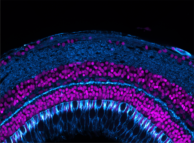
Figure 6: High-resolution multi-fields of view image of a zebrafish retina labelled with 2 channels - Phalloidin (cyan) and DAPI (magenta). Image acquired with a Andor confocal microscope system and Andor sCMOS camera.
Summary of Key Differences
| Parameter | Monochrome camera | Colour camera | Practical impact |
| Sensitivity | Captures more usable light because there is no Bayer filter array. | Colour filters block a proportion of incoming photons at each pixel. | Monochrome cameras improve signal-to-noise ratio, reduce exposure times and help limit photobleaching and phototoxicity in fluorescence experiments. |
| Spatial resolution | Samples intensity directly at every pixel. | Samples each colour at only a subset of pixels and reconstructs the image by interpolation. | Monochrome cameras preserve fine structural detail more effectively, while colour cameras can have lower effective resolution for small features. |
| Colour information | Requires filters as part of the microscope light path, or pseudo-colour assignment to build colour or multichannel images. | Provides direct RGB images in a single exposure. | Colour cameras are convenient when natural colour appearance carries diagnostic or classification value. |
| Quantitative imaging | Records intensity directly at every pixel without colour interpolation. | Demosaicing and colour filter cross-talk can introduce artefacts and reduce measurement accuracy. | Monochrome cameras are generally preferred for measuring fluorescence intensity, weak signals and small changes over time. |
| Cost | Wide price range – scientific-grade cameras will cost considerably more than basic models. | Often low-cost as used in less demanding applications where signal fidelity is not a factor. | Monochrome cameras are often much higher cost than colour cameras as the overall camera design is more developed, e.g. more sophisticated electronic design and thermoelectric cooling. |
Application Guidance
- Choose a colour camera when the sample is brightly illuminated and natural colour reproduction is important. Common examples include H&E-stained tissue, chromogenic assays and teaching demonstrations. Colour cameras simplify acquisition because the user can capture a complete colour image without changing filters or merging channels.
- Choose a monochrome camera when sensitivity, resolution or quantitative accuracy matters most. A modern sCMOS like a ZL41 Cell or Sona camera is generally the best option for fluorescence microscopy, weak transmitted-light, high-speed imaging, time-lapse experiments and any application where photon budget is limited. For very challenging low light applications like single molecule biophysics, an iXon EMCCD camera would be recommended.
Conclusion
Colour cameras are low cost yet effective for immediate, accurate-looking colour images to support interpretation, documentation or classification. Monochrome cameras carry a higher price but deliver clear advantages when experiments depend on capturing the maximum number of photons and measuring signal reliably. For brightfield and stained-sample workflows, colour cameras are often the practical choice. For fluorescence and quantitative scientific imaging, where signal levels are low and accurate quantification is critical, monochrome cameras are the preferred technical solution.
Recommended Monochrome Camera Options
- ZL41 Cell: The ideal solution for routine fluorescence microscopy offering flexible performance at a relatively low price.
- 4.2 & 5.5 Megapixel formats with 6.5µm pixel size
- Up to 100 fps
- Up to 82% QE
- Sona-6 Extreme: Excellent detection of weak signals at high speed.
- peak QE of 95%
- Very low 1.0e- read noise
- Up to 135 fps full frame
- CB2 High Res: Exceptional high speeds and high resolutions at low magnifications.
- 24.5-megapixel camera with Global Shutter technology
- Up to 386 fps
- on-chip binning and other features for imaging dynamic processes
- iXon EMCCD: The gold standard in EMCCD detection for biophysics and single molecule studies.
- Single photon sensitivity
- Back-illuminated >95% QE
- Fast frame rates

