|
|
|
|
RISE Microscopy is a novel correlative microscopy technique that combines SEM and confocal Raman Imaging. Through RISE Microscopy ultra-structural surface properties can be linked to molecular compound information.
The RISE Microscope combines all features of a stand-alone SEM and the confocal Raman imaging microscope witec360 within one instrument:
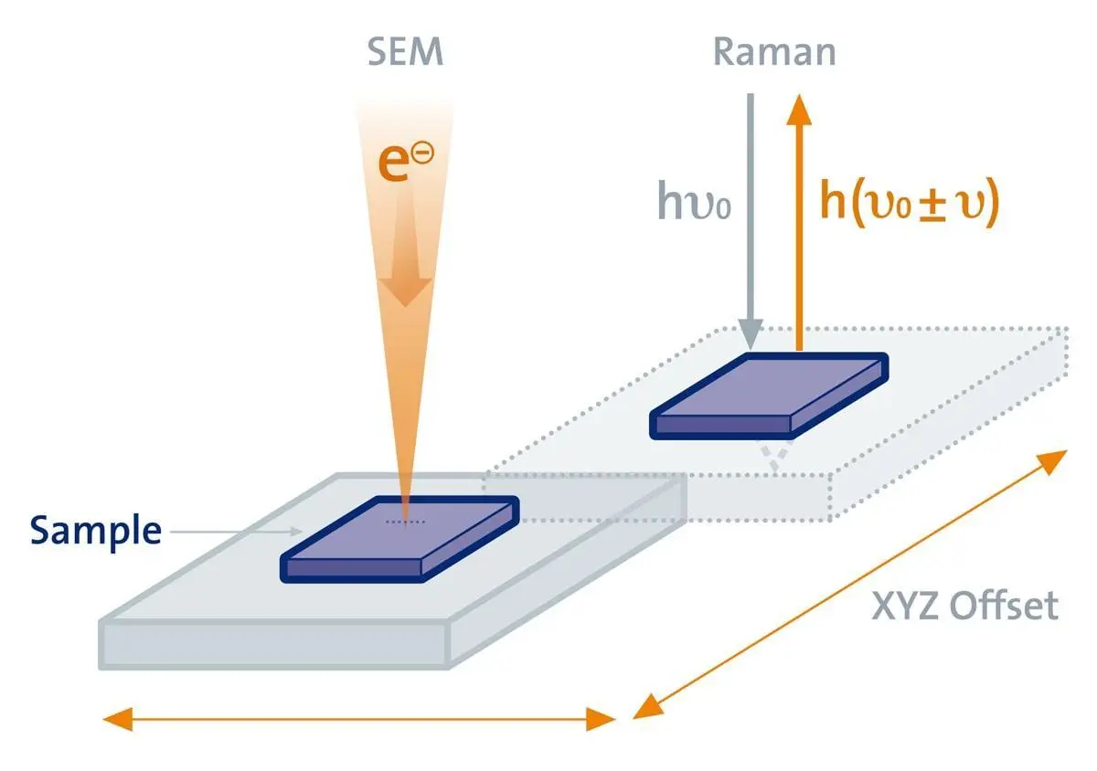
Samples are automatically transferred from one measuring position to the other within the vacuum chamber of the combined Raman-SEM instrument, streamlining the workflow and drastically improving the instrument's ease of use.
See more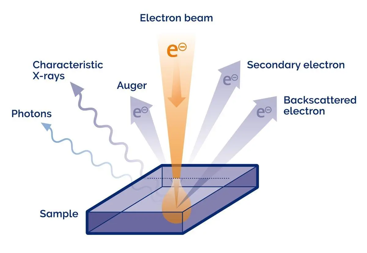
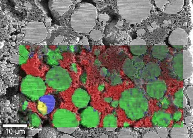
RISE (Raman-SEM) image of the cathode of a fast cycled lithium-ion battery. Changes due to repeated cycling influence the composition of the lithium metal oxide particles. Sample courtesy of Dean Miller (Tescan USA)
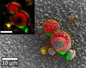
RISE (Raman-SEM) image of electrochemically deposited Li(NiMnCo)O2 battery particles. Different colors indicate different local cobalt and nickel concentrations. The inset shows the Raman image. The scale bar indicates 10 µm in both pictures. Sample courtesy of Timo Sörgel, Gerhard Schneider (Institut für Materialforschung, Hochschule Aalen, Germany)
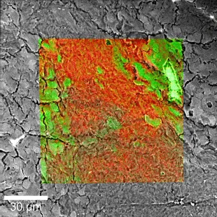
Raman-SEM image overlay of a hamster brain tissue sample. Color-coded Raman image parameters: Green: White brain matter; Red: Gray brain matter; 100 µm x 100 µm, 300 x 300 pixels = 90,000 spectra, 50 ms integration time per spectrum.
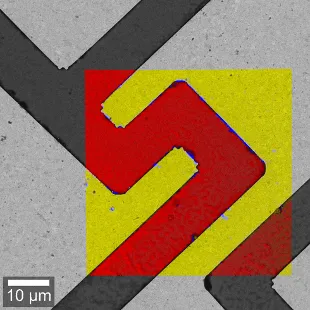
Raman-SEM image overlay of a GaAs semiconductor sample. Color-coded Raman image parameters: Yellow: Gold substrate; Red: GaAs; Blue: Residues from production; 50 µm x 50 µm, 300 x 300 pixels = 90,000 spectra, 34 ms integartion time per spectrum.
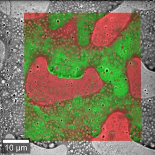
Raman-SEM image overlay of a PMMA-PS polymer blend. Color-coded Raman image parameters: Green: Polystyrene; Red: Polymethyl methacrylate.
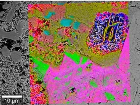
Raman-SEM image overlay of a piece of hematite (Fe2 O3). SEM reveals structural characteristics, but cannot differentiate between the different oxides present in the hematite. However, the Raman spectra indicate the occurrence of several crystal forms of hematite and of goethite (FeO(OH)).
Our experts demonstrate how RISE Microscopy can be applied across a range of research areas, showcasing its capabilities and practical applications.
Explore the video clips below to discover RISE Microscopy in action. All videos are available with localised captions in English, Traditional Chinese and Korean.
This tutorial will provide an overview of the RISE microscope at the Oxford Instruments Ulm Site. Starting with SEM imaging, we will analyze porous SiC ceramics consisting of various polytypes of SiC. These SiC polytypes can be identified with Raman imaging by analyzing the molecular bonds within the crystal structure. The correlation between Raman and SEM images will be highlighted.
You will learn:
Due to their unique optical and electronic properties, two-dimensional (2D) materials have great potential for application in optoelectronic devices, sensors energy storage, catalysis and many more. Diverse crystal properties such as the number of layers and growth defects influence the optical and electronic properties of the 2D materials. The aim of this tutorial is to show how RISE contributes to the analysis of 2D materials.
The development and production of drug delivery systems requires efficient and reliable control mechanisms to ensure the quality of the final products. These products can vary widely in composition and application. Therefore, analytical tools such as RISE Microscopy are essential for investigating pharmaceutical materials including powders and tablets.
Over the years biominerals have been refined by natural selection, leading to the unique forms and functions we observe today. These forms can be studied at high resolution using the SEM. The association of such structures with changes in chemical bonds leads to a deeper understanding of processes. In this session the fine structure observed on nacre with an SEM will be correlated to anisotropies within the crystal structure of CaCO3
Scanning electron microscopy is a powerful tool for the analysis of geological samples. In combination with EDS, the high-resolution SEM images can be complemented with elemental composition of various mineral phases. Often however this does not lead to the complete picture. Raman spectroscopy and imaging provide additional information about the molecular bonds, thus can distinguish polymorphs and polytypes.
If you'd like to learn more about the possibilities of Raman imaging for your individual field of application, our specialists will be happy to discuss them with you.

© Oxford Instruments 2026