Jun. 30, 2021
Photon antibunching identifies single-photon emitters
WITec combines antibunching experiments with fast Raman and photoluminescence imaging.
Single-photon emitters have quantum mechanical properties that are exploited in quantum technology and information science, including the development of quantum computers and cryptography methods. Nitrogen vacancy (NV) centers in diamonds, single fluorescent molecules, carbon nanotubes and quantum dots are prominent examples of single-photon emitters. In order to identify them in a sample, antibunching experiments are commonly performed.
Antibunching is a quantum mechanical effect that reveals the particle-like behavior of light. It arises because a single-photon emitter can only emit one photon at a time. The minimum interval between photon emissions depends primarily on the excited-state lifetime of the emitter, because a cycle of excitation and relaxation must be completed between two photons. If the signal is split and measured with two detectors, each single photon can only be detected by one of them. Antibunching therefore results in an anticorrelation of the two detectors’ signals at very short lag times (Hanbury Brown-Twiss experiment).
Here WITec in cooperation with PicoQuant demonstrates the integration of antibunching measurements within a confocal Raman microscope. This combination makes it possible to characterize a sample with fast Raman and photoluminescence (PL) imaging and identify areas of interest for subsequent antibunching experiments with the same instrument, a WITec alpha300 Raman microscope. Antibunching measurements are performed in a Hanbury Brown-Twiss configuration, where the signal is split by a 50/50 beam splitter and detected by two APDs. Both detectors are connected to a MultiHarp 150 time-correlated single photon counting (TCSPC) unit from PicoQuant, which records the delay between two single-photon events at picosecond resolution. A histogram of the time differences shows a pronounced dip for very short times, i.e. antibunching, if the investigated structure is a single-photon emitter. Lifetime measurements are additionally possible in this configuration. A 532 nm continuous wave laser was applied for excitation here, but the setup also supports other wavelengths and pulsed laser sources.
We demonstrate this functionality using a sample of diamond micropillars, a fraction of which contain single NV centers. The sample was provided courtesy of Dr. Rainer Stöhr and Prof. Dr. Jörg Wrachtrup from the 3rd Physics Institute at the University of Stuttgart, Germany.
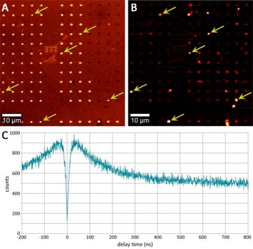
The pillars were first imaged with Raman and PL microscopy. The Raman image represents the intensity of the diamond peak at 1330 cm-1 and reveals the positions of intact pillars (Fig. A). In the fluorescence image, some pillars are particularly bright, indicating the presence of NV centers (Fig. B). By comparing the Raman and PL images, structures of interest can be distinguished from fluorescent contaminations on the sample: intact pillars with NV centers exhibit a strong diamond Raman signal and bright fluorescence (arrows in Fig. A and B), while contaminations lack the Raman signal.
Antibunching experiments were performed at some of the identified structures of interest in order to test for the presence of single NV centers. The resulting correlation curve for one selected pillar is displayed in Fig. C. The histogram has a pronounced dip at a detection time difference of zero. This indicates that the observed micropillar indeed contained a single NV center and was a single-photon emitter. The observed drop in the curve toward longer delay times reveals the presence of a shelving state, which is a well-known phenomenon for diamond NV centers.
The integration of antibunching experiments within a confocal Raman microscope offers many benefits. Such an instrument is capable of carrying out both spatially resolved chemical characterization and quantum mechanical investigations. As demonstrated here, the correlation of Raman and photoluminescence signals can pre-select candidate locations for NV centers to be subsequently confirmed by antibunching experiments. This provides valuable insight and an accelerated workflow to researchers exploring single photon emitters for use in emerging technologies, including quantum computers.