

1 Nov
Andor Launches New ZL41 Cell and Sona sCMOS Cameras
Easy and Cost-Effective Super-Resolution to ~100 nm
Andor Technology, an Oxford Instruments company and a world leader in scientific imaging solutions, has today announced the launch of two new scientific CMOS cameras, specifically designed for life science researchers.
These new product introductions further strengthen Andor’s broad portfolio of cameras and microscopy systems, while the company also offers market-leading image analysis software with Imaris.
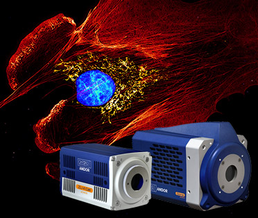
New ZL41 Cell sCMOS: Performance and Flexibility
ZL41 Cell is the next generation in the highly successful Zyla sCMOS series. With ZL41 Cell, it is possible to transform a normal fluorescence microscope into a super-resolution microscope using our exclusive SRRF-Stream+ technology. ZL41 Cell offers remarkable flexibility, as well as superb sensitivity, speed and resolution, making it ideal for those that are upgrading their imaging system, or for those that have specific application needs such as live-cell imaging, or Single Molecule Localisation Microscopy.
Dr. Alan Mullan, Andor's Product Manager for Microscopy Cameras, commented:
"The combination of the ZL41 Cell and SRRF-Stream+ opens up super-resolution capabilities to a much wider range of researchers than ever before, and at a much lower price point. You can use your existing labelling protocols, and normal fluorescence microscope to obtain highly detailed and vibrant images in real-time. Therefore, super-resolution to 100 nm becomes accessible even to those that previously considered the technique to be difficult or costly.”
Sona Back-illuminated sCMOS: Now Even Faster and More Sensitive
The Sona-6 back-illuminated sCMOS has been enhanced to push the limits of detection further, even for the most challenging, light starved imaging applications such as live-cell confocal and single molecule studies. The noise floor has been reduced to ≤1.0e-, which, when combined with the 95% Quantum Efficiency and lowest dark current, allows detection of the weakest signals. This means exposures and phototoxicity are reduced, maintaining cell physiology, for fast and gentle imaging of delicate cells over longer durations.
A new High-Speed mode has been implemented to meet the needs of fast imaging applications like calcium and ion imaging, FRET and blood flow studies. By exploiting a 2-lane CoaXPress connection, 135 fps of sustained and stable high-speed operation is now possible. Further optimizations have enhanced image quality for even clearer and more vibrant images.
Dr. Alan Mullan stated:
“Typically, running sensors really fast increases the noise floor significantly while dynamic range and the ability to make quantitative measurements decreases. This can make it difficult to get the benefit of the high-speed capabilities in all but a few limited cases. With the Sona-6 we set out to boost speeds, but crucially still maintain a high signal to noise, a suitable dynamic range and quantitative accuracy for applications that require precision and accuracy as well as outright speeds. The result of this is that the updated Sona-6 is well suited to a wide range of applications that demand high temporal resolution.”
Dr. Leonidas Asimakoulas, Andor’s Physicist and Optical Lead for the Sona development project added:
“Sona has proven to be a valuable research tool for many researchers across life sciences. By incorporating new features and pushing the GS2020BSI sensor performance to the maximum, researchers will be able to benefit from the lower noise floor and image at higher speeds than was previously possible to help answer their research questions.”
The latest updates to Sona-6 add to an already impressively capable and flexible imaging camera that is sure to help many researchers push our understanding of many fields of research across life sciences even further.
Image Caption: Revealing the cytoskeleton of the cell in highly-resolved vibrant detail with Andor’s ZL41 cell and SRRF-Stream+ super-resolution technology. Actin (Red), Mitochondria (Yellow), Nucleus (Cyan). Credit: Motosuke Tsutsumi, National Institute of Physiological Science.
