FAQs & Glossaries
What is Nuclear Magnetic Resonance? Common Questions About NMR
Author: Judith Beer
Published: 02 Nov 2026 · Last updated: 02 Nov 2026
Introduction
Nuclear magnetic resonance (NMR) spectroscopy is an analytical technique used in academic research and industry to study the chemical composition, structure and physical properties of samples. NMR has for a long time been associated with specialist high-field instruments. However, time-domain NMR analysers and compact benchtop NMR spectrometers have made routine NMR analysis possible in standard laboratories.
This FAQ introduces the basic principles of nuclear magnetic resonance (NMR), common NMR experiments, applications, and practical aspects of NMR analysis.
What is NMR?
Nuclear magnetic resonance (NMR) is a physical effect that occurs when atomic nuclei with specific magnetic properties respond to a magnetic field. The term NMR also describes the analytical methods that measure this response.
NMR can provide information about a sample’s chemical identity, composition, structure, and physical properties. The information obtained depends on the NMR technique and the way the NMR signal is measured and analysed.
How does NMR work?
NMR uses the magnetic properties of certain atomic nuclei to probe their chemical and physical environments. The sample is placed in a magnetic field, which creates a net alignment of the atom’s nuclear spins. Short radio-frequency pulses disturb this alignment and cause the nuclear spins to transition to higher energy levels.
As the nuclei return to equilibrium, they generate an NMR signal visible in the time domain. The rate at which the signal returns towards equilibrium is described by the relaxation time.
In NMR spectroscopy, the detected time domain signal, often called the free induction decay (FID), is converted into an NMR spectrum using a Fourier transformation. The position and intensity of signals depend on the nuclei and their local molecular environments. Analysing the resulting NMR spectra can provide structural information and determine the concentration of components in a sample.
Alternatively, the time domain NMR signal can be analysed directly to obtain information on phase distribution molecular mobilities and other physical properties of the material.
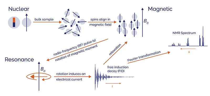
Schematic overview of how NMR spectroscopy works
What is NMR used for?
NMR is used to identify substances, determine molecular structures, measure composition, analyse formulations, verify raw materials, and assess sample purity. Applications range from research on small molecules and materials to routine industrial analysis.
NMR is frequently used in the chemical, pharmaceutical, food, energy, polymer, and materials sectors. Industrial applications include process monitoring, quality assurance, and quality control.
What is TD-NMR?
Time-Domain NMR (TD-NMR), also known as NMR relaxometry, measures the relaxation and diffusion behaviour of nuclear spins over time. Differences in these properties can be used to quantify components such as water, oil, and solid fat and to distinguish between phases or states in a sample, e.g. amorphous and crystalline content.
TD-NMR provides fast, robust and non-destructive measurements and can analyse even large or heterogeneous samples. It is used for both, research and quality control in areas including agriculture, food, polymers, petrochemicals, and materials science.
Unlike NMR spectroscopy, TD-NMR uses less homogeneous and lower field strength magnets. It therefore does not resolve individual chemical environments in the same way as NMR spectroscopy. Instead, TD-NMR analyses the time-domain NMR signal directly to study properties such as composition, molecular mobility, phase distribution, pore structure, particle or droplet size, and diffusion.
Learn more about TD-NMR

What is benchtop NMR spectroscopy?
Benchtop NMR spectroscopy uses compact, cryogen-free NMR instruments with permanent magnets instead of superconducting magnets. This gives the instruments a smaller footprint and allows them to operate in standard laboratory and manufacturing environments.
Benchtop NMR spectroscopy operates at lower magnetic field strengths than conventional high-field NMR, typically in the range of 1.0 – 2.1 T. Depending on the instrument, it can support experiments including one-dimensional and two-dimensional NMR, gradient-based measurements, and measurements of different NMR-active nuclei (broadband NMR).
Learn more about benchtop NMR
Which nuclei can NMR detect?
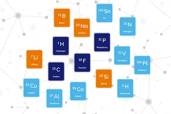
NMR can detect nuclei with non-zero nuclear spin, often described as NMR-active nuclei. These nuclei have a magnetic moment that interacts with the magnetic field of an NMR instruments.
Common nuclei measured by NMR spectroscopy include hydrogen-1 (1H), carbon-13 (13C), fluorine-19 (19F), and phosphorus-31 (31P). Whether other nuclei can be detected depends on factors including natural abundance, sensitivity, linewidth, resonance frequency, and the instrument configuration.
TD-NMR most commonly measures 1H or 19F because of their high natural abundance and sensitivity. These nuclei can be used for measuring bulk properties of water, oil, fluorine, and solid fat.
Can NMR be used for quantitative analysis?
Yes. NMR can provide relative and absolute quantitative measurements, commonly called quantitative NMR or qNMR. Under suitable acquisition conditions, NMR signal intensity, measured from the signal area, is proportional to the number of contributing nuclei.
Relative qNMR compares signals from components in the same sample. Absolute qNMR determines the amount or concentration of an analyte by comparison with an internal or external standard of known quantity or concentration.
Accurate quantitative measurements require suitable sample preparation, relaxation delays, signal selection, and data processing.
How long does an NMR measurement take?
An NMR measurement can take from seconds to hours, depending on the experiment, sample, nucleus, and required signal quality. Routine TD-NMR measurements commonly take seconds to a few minutes. Simple one-dimensional NMR spectra can also be acquired quickly when the sample concentration and nucleus NMR sensitivity are sufficient.
Longer measurements may be needed for dilute samples, less sensitive nuclei, quantitative experiments with long relaxation delays, or multi-dimensional NMR experiments. Acquisition time also depends on sample volume, number of scans, magnetic field strength, and the required signal-to-noise ratio.
How do you read an NMR spectrum?
Reading an NMR spectrum involves examining chemical shift, splitting patterns, and signal integration together.
- Locate the chemical shifts: The chemical shift along the horizontal axis indicates the local chemical environment of the observed nucleus. Peak positions are reported in parts per million (ppm).
- Compare integration values: The area under a signal is related to the number of contributing nuclei. For example, an integral twice as large as another can indicate twice as many corresponding nuclei.
- Analyse splitting patterns: Multiplets result from spin-spin coupling of neighbouring nuclei and can provide information about molecular connectivity.
These features should be interpreted together, as peak overlap, exchange, solvent signals, and sample conditions can affect the appearance of an NMR spectrum and impact individual observations.
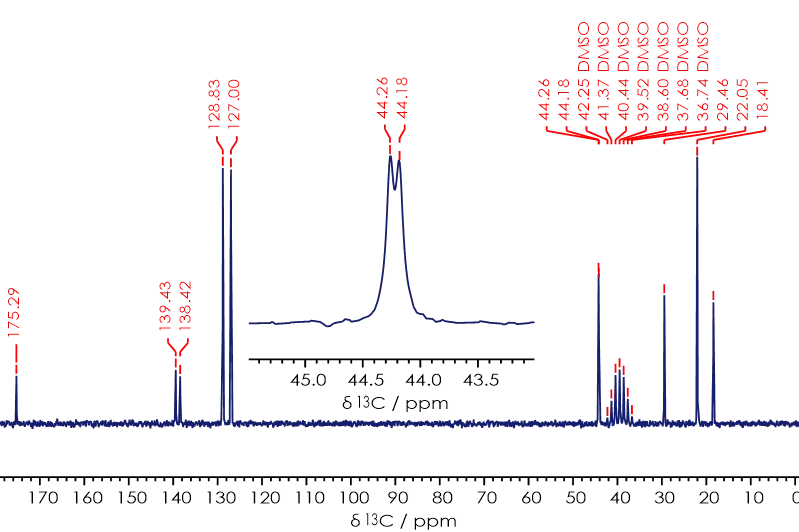
13C{1H} NMR spectrum of Ibuprofen
What causes chemical shift in NMR?
Chemical shift is caused by differences in the local electronic environment around an observed nucleus. Electrons respond to the applied magnetic field and create a small local field that partly shields the nucleus.
The amount of shielding depends on factors such as neighbouring atoms, bonding, functional groups, solvent, concentration, and temperature. Nuclei in different environments therefore resonate at different frequencies.
Chemical shift describes this frequency difference relative to the Larmor frequency of the observed nucleus. It is reported in parts per million (ppm), which makes chemical-shift values independent of magnetic field strength and allows comparison of spectra acquired on different NMR instruments at different magnetic field strengths.
How do you determine molecular structure using NMR spectroscopy?
NMR spectroscopy determines molecular structure by combining information from chemical shifts, signal integrals, splitting patterns, and coupling constants from different measurements. The analysis is often iterative: each observation helps refine the proposed molecular structure until it is consistent with the available NMR data.
One-dimensional 1H and 13C NMR spectra can identify distinct chemical environments and suggest functional groups. J-coupling and multiplet patterns provide additional information about connections between nuclei, such as how many bonds separate interacting nuclei.
Multi-dimensional experiments such as COSY or HSQC show correlations between nuclei and help establish molecular connectivity. Measurements of other nuclei, such as for 19F or 31P, can provide additional evidence where relevant.
Learn more in our Application Note Structural Elucidation of Ibuprofen
What is 1H NMR?
1H NMR, or proton NMR, measures signals from hydrogen-1 nuclei in a sample. It provides information about chemical environments, neighbouring nuclei, and the relative numbers of hydrogen atoms through chemical shifts, coupling patterns, and integrals.
Hydrogen is present in most organic compounds and gives relatively strong signals, even at short acquisition times. For this reason, 1H NMR is the most common NMR technique used to identify small molecules and determine molecular structure.
Complex samples can contain overlapping peaks that make interpretation more difficult. In these cases, 1H NMR can be combined with 13C NMR or two-dimensional NMR experiments.
What is 13C NMR?
13C NMR measures carbon-13 nuclei and shows the different carbon environments in a sample. 13C signals occur across a broad chemical shift range of around 0 to 220 ppm, which can help distinguish different functional groups and reduce overlap with 1H NMR.
13C has a natural abundance of approximately 1.1 %, so its signals are less sensitive than 1H signals. Measurements therefore commonly require more scans and longer acquisition times.
What is J-coupling and how is it calculated?
J-coupling is an effect that arises from through-bond interactions between different NMR active nuclei. It can help establish bond connectivity and stereochemical relationships.
J-coupling is determined by measuring the separation (in Hz) between peaks in a multiplet. The magnitude of J-coupling constants are independent of the magnetic field strength and can range from 6 to 18 Hz for a three-bond 1H-1H coupling, to hundreds of Hz for coupling between heteronuclear.
Key takeaways
- Nuclear magnetic resonance (NMR) describes both a physical phenomenon and the analytical techniques based on it.
- NMR spectroscopy uses frequency-domain spectra for chemical identification, structural analysis, and quantification.
- TD-NMR analyses time-domain signals directly to study composition, relaxation, diffusion, molecular mobility, and phase-related properties.
- The sample, nucleus, analytical question, NMR instrument, and required signal-to-noise ratio influence the choice of an NMR experiment.
- Chemical shift, coupling, and signal integration provide complementary information for interpreting NMR spectra.