Introduction
NMR spectroscopy is most commonly used to analyse hydrogen-1 (1H, proton), carbon-13 (13C) and fluorine-19 (19F) nuclei. However, most modern NMR spectrometers can observe many more nuclei than that. The Oxford Instruments X-Pulse 90 MHz & 60 MHz Benchtop NMR spectrometers are fully tunable broadband instruments, able to observe a wide range of nuclei on a single instrument.
This article introduces the X-Pulse's broadband NMR capabilities by showcasing a series of example spectra acquired of various X-nuclei containing compounds. It highlights important features when acquiring NMR spectra for the different nuclei.
X-Pulse Broadband configuration
Like most NMR spectrometers, the X-Pulse is a three channel spectrometer: One channel is optimised for a deuterium (2H, hydrogen-2) lock signal; another one for proton and fluorine-19 (19F) signals; and the third, broadband X-channel, is suited for a wide range of other nuclei, often referred to as X-nuclei. In its standard broadband configuration, the X-channel covers a frequency range between 41% and 19% that of the proton. The periodic tables shown in Figure 1, demonstrate the range of nuclei possible to observe on a X-Pulse 90 or X-Pulse 60 in its standard broadband configuration.
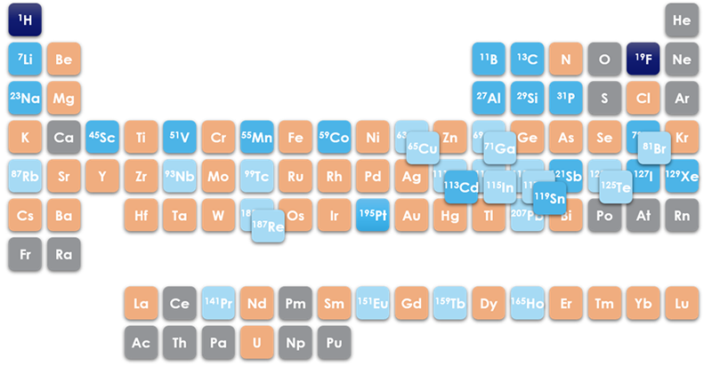
Figure 1 Periodic Table of the Elements, showing nuclei observable on the X-Pulse; dark blue = 1H/19F, blue = nuclei observed on the X-channel (at the time of writing); light blue = within frequency range of X-channel; orange = no NMR active nuclei within range; grey = no NMR active nuclei with >1% natural abundance.
Carbon-13 NMR Spectroscopy
Carbon-13 (13C) is likely the second most studied nucleus by NMR spectroscopy, after protons, due to the importance of carbon in organic chemistry and the life sciences. However, due to its low natural abundance and gyromagnetic ratio, it has one of the lowest receptivity values of any NMR active nuclei. Indeed, if not for its ubiquitous nature, 13C NMR would likely be an uncommonly studied nucleus by NMR spectroscopy, rather than one of the driving forces in its development. Instead, NMR spectroscopists have developed many useful techniques to examine this crucial element, such as advanced two-dimensional proton-carbon correlation experiments.1
13C NMR spectra are usually obtained using a technique known as decoupling, to remove the proton-carbon couplings, which greatly simplifies the spectrum and enhances the signal-to-noise ratio. Most small organic molecules typically produce proton-decoupled 13C (commonly denoted: 13C{1H}) NMR spectra made up of a series of sharp singlets over the chemical shift range of δC 0 to +200 ppm. Unlike in 1H and 19F NMR spectra, the peak area of a signal in a standard 13C NMR spectrum will not typically correlate with the number of carbon atoms in the sample, making the spectra more useful for qualitative, rather than quantitative, applications.
A 13C NMR spectrum of ethyl crotonate (Figure 2), for example, clearly distinguishes the different carbon environments. The spectrum shows the carbonyl carbon at δC 166 ppm, the alkene carbons in the range δC 120 - 145 ppm, the ether-adjacent carbon at δC 60 ppm, and the methyl carbons between δC 10 - 20 ppm.
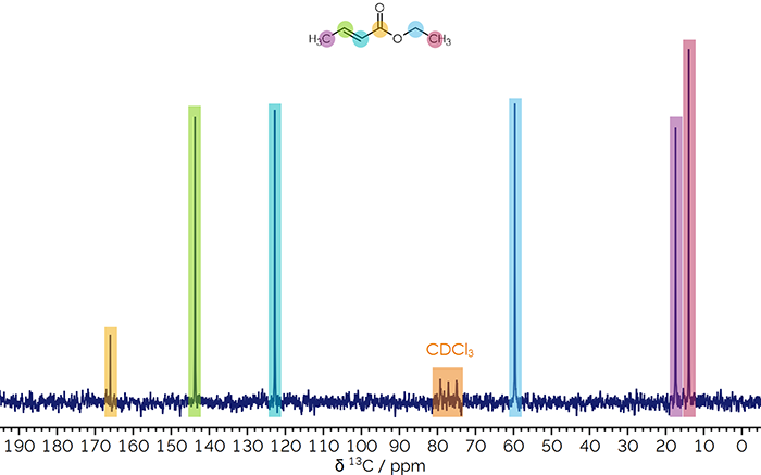
Figure 2 13C{1H} NMR spectrum of ethyl crotonate in CDCl3.
1Read more about two-dimensional experiments here
Phosphorus-31 NMR Spectroscopy
Phosphorus-31 (31P) NMR spectroscopy is commonly used in coordination chemistry, and hence catalysis, where phosphorus containing ligands are often employed (for example those shown in Figure 3a). Also, due to the importance of phosphate (PO43−) in many biochemical pathways, phosphorous containing compounds are found in pharmaceuticals and dietary supplements which are amenable to study by NMR. Additionally, there are many phosphorus containing anions, for example hexafluorophosphate (Figure 3a), which is commonly used in lithium-ion batteries. As with 13C NMR spectra, 31P spectra are commonly proton decoupled, giving generally sharp signals over a wide range of δP −180 to +250 ppm.
Due both to the wide chemical shift range and the fact that most phosphorus containing compounds have only a few phosphorus atoms, 31P NMR spectroscopy is ideal for distinguishing them. For example, triphenylphosphine {PPh3}, triphenylphosphine oxide {OPPh3}, triphenyl-phosphite {P(OPh)3}, and triphenylphosphate {OP(OPh)3}, have effectively identical 1H and 13C NMR spectra arising from the phenyl rings, yet can be readily distinguished by their 31P{1H} NMR spectra (Figure 3b).2
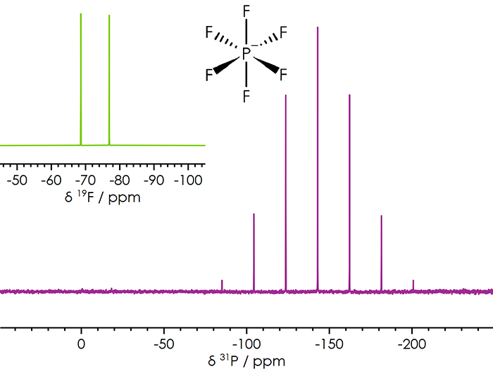
Figure 3a 31P NMR spectrum of the hexafluorophosphate anion; insert of the corresponding 19F NMR spectrum; 1JPF = 706 Hz.
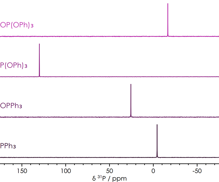
Figure 3b 31P{1H} NMR spectra of triphenylphosphine, triphenylphosphine oxide, triphenylphosphite, and triphenylphosphate.
2For more examples, see here
Boron-11 NMR Spectroscopy
Unlike 1H, 13C and 31P, boron-11 (11B) is a quadrupolar nucleus (which means that it has a nuclear spin greater than 1/2), leading to different NMR properties. The 11B spin of 3/2 leads to generally broader signals than spin 1/2 nuclei, which are observed over a range of δB −120 to +90 ppm. Boron containing compounds have a range of applications, including as Lewis acid catalysts and in Suzuki cross-coupling reactions (Nobel Prize in Chemistry 2010); hence, they are commonly used as intermediates in the synthesis of fine chemicals and pharmaceuticals.
One complication with 11B NMR spectroscopy is that the glass commonly used to make NMR tubes and sometimes components of the NMR spectrometer contains boron, often leading to a background signal in the NMR spectrum. This background signal can be minimised and even eliminated by appropriate hardware and experimental design. As demonstrated in Figure 4a, while a background signal (causing a wide, flat hump in the baseline) is present in the spectrum obtained on a high-field instrument, none is visible in the spectrum collected on the X-Pulse.
Since 11B is a quadrupolar nucleus, the linewidths of its NMR signals are strongly dependent on the size of the molecule and symmetry around the boron centre. For example, the four-coordinate tetrahedral tetra(aryl)borane shown in Figure 4a has a peak width of 20 Hz; while the three-coordinate trigonal-planar boronic acid derivative shown in Figure 4b, has a peak width greater than 200 Hz; and the small tetrahedral tetrafluoroborate anion in Figure 4c has peak widths of less than 1 Hz. As a result, simple linewidth measurements of a boron spectrum can be a probe of structure and symmetry.
The spectra of the tetrafluoroborate anion in Figure 4c demonstrate an important feature of quadrupolar nuclei in NMR spectroscopy. Most NMR courses teach the "n+1 rule" to determine the number of peaks in a multiplet: "if a nucleus has n equivalent nearest neighbours, they will split the signal into a multiplet consisting of n+1 peaks", with the peak intensities predicted using Pascal's triangle. However, this rule is valid only for spin 1/2 nuclei such as 1H, 13C, and 19F. As a result, while the 11B spectrum shows a 1:4:6:4:1 pentet arising from coupling to four equivalent spin 1/2 19F nuclei, the corresponding 19F spectrum (inset, Figure 4c) shows a 1:1:1:1 quartet, due to coupling to a single spin 3/2 11B nucleus. In contrast, coupling to a single spin 1/2 nucleus would produce a 1:1 doublet (see for [PF6]−, Figure 3a, insert).
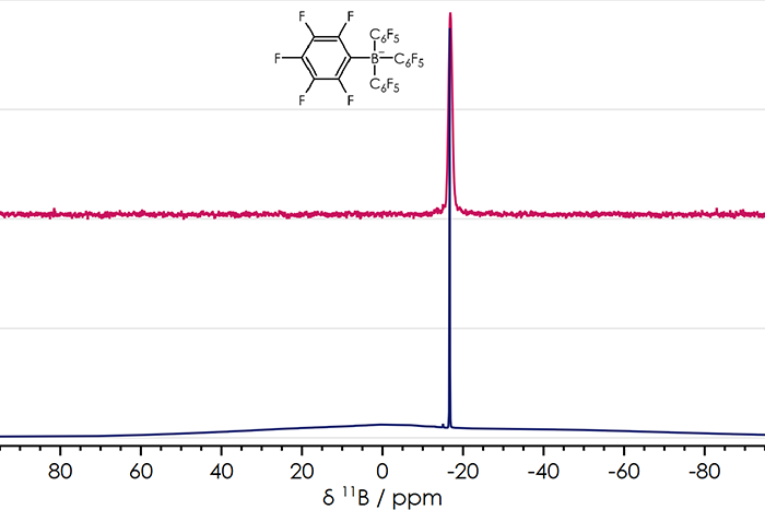
Figure 4a 11B NMR spectra of a tetra(aryl)borane recorded on an X-Pulse 60 (cerise, top) and a high-field instrument (blue, bottom); peak widths at half height are 20 Hz.
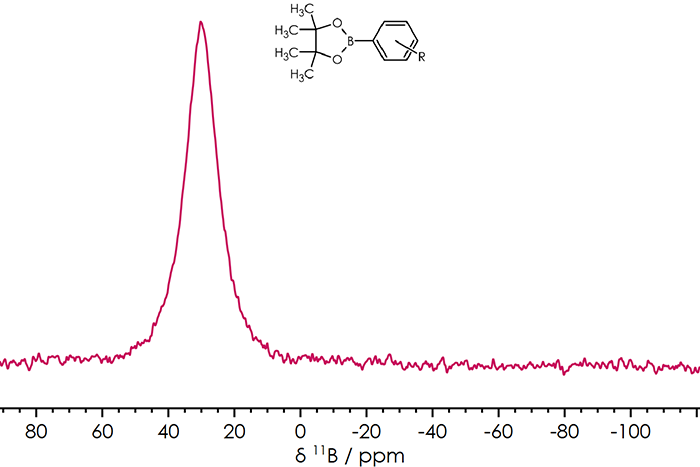
Figure 4b 11B{1H} NMR spectrum of phenylboronic acid pinacol ester on an X-Pulse 60; peak width at half height is > 200 Hz.
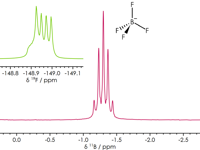
Figure 4c 11B NMR spectrum of tetrafluoroborate in aqueous solution acquired on an X-Pulse 90; the inset shows the corresponding 19F NMR spectrum; peak widths at half height are < 1 Hz; 1JBF = 2 Hz
Sodium-23 & Lithium-7 NMR Spectroscopy
Sodium is the sixth most abundant element on earth and is ubiquitous in living organisms. In addition, sodium containing compounds are valuable reagents in organometallic chemistry. Moreover, sodium is a component of an extremely wide range of substances from food and drink to pharmaceuticals, petroleum additives, polymers, and next-generation sodium-ion batteries just to name a few.
As with 11B, sodium-23 (23Na) is a quadrupolar nucleus, with a linewidth strongly dependent on the local symmetry and the size of the molecule. 23Na signals can be observed over the range of δNa +10 to −60 ppm. Sodium cations in aqueous solutions give sharp signals close to 0 ppm (Figure 5).
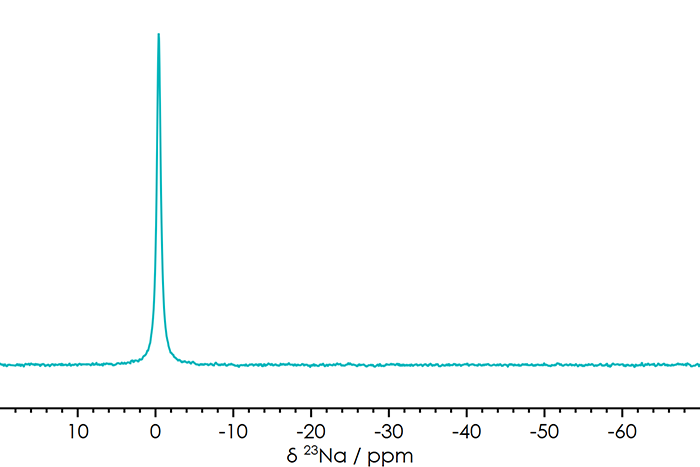
Figure 5 23Na NMR spectrum of Na+ in aqueous solution acquired on an X-Pulse 90; peak width at half height is 9 Hz.
As with sodium, lithium containing compounds are valuable reagents in organometallic chemistry. Unlike sodium, lithium does not have a known biological role, although it is used in pharmaceuticals. One important application of lithium is in energy storage systems, with solutions containing Li+ cations, such as those used in batteries, are amenable for study by NMR spectroscopy.
As with 11B and 23Na, lithium-7 (7Li) is a quadrupolar nucleus with linewidths strongly dependent on molecular symmetry and size. 7Li signals appear over a range from δLi +15 to −20 ppm. Like sodium, lithium cations in aqueous solutions give sharp signals near 0 ppm.
Figure 6 shows a series of Pulsed Field Gradient Spin-Echo (PGSE) NMR spectra of the lithium cation in a dialkyl carbonate, as is commonly used in lithium-ion batteries. By obtaining these spectra at varying gradient strengths (between 0 and 36 G/cm), the diffusion coefficient of Li+ may be directly measured. The knowledge of diffusion coefficients is valuable for the analysis of electrochemical data, and hence in the development of batteries and super-capacitors.3
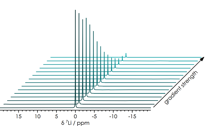
Figure 6 Series of 7Li PGSE NMR spectra of Li+ in alkyl carbonates acquired on an X-Pulse 90, for measuring diffusion coefficients in battery electrolytes, with the applied gradient strengths varying from 0 to 39 G/cm.
3More detail about the use of NMR diffusion measurements on battery electrolyte can be found here
Silicon-29 NMR Spectroscopy
Silicon is another extremely abundant element with many industrial uses, including being a major component of computers and electronics, and as a major constituent of glass. While some of silicon's most familiar applications are in a solid form, silicon-29 (29Si) benchtop NMR spectroscopy is useful for other applications, such as the analysis of poly-siloxanes (silicones) and their monomer precursors, which have a range of applications including as adhesives, lubricants, and sealants, along with uses in medicine and in the home.
Like 13C, 29Si has a low receptivity due to both its low natural abundance and gyromagnetic ratio. Spectra are usually obtained using inverse-gated proton decoupling, giving rise to sharp signals while minimising reduction of the 29Si signal that can occur from other decoupling methods. Signals appear over the very wide range from δSi −350 to +175 ppm. Some of the methods for sensitivity enhancement used in 13C NMR are also applicable to 29Si NMR. Examples include the use of polarisation-transfer, and two-dimensional pulse sequences, where it's the higher receptivity protons, rather than the X-nuclei, which are excited.
The simple one-dimensional spectrum shown in Figure 7a shows two signals, one for each of the two silicon environments, while the HMBC two-dimensional correlation spectrum in Figure 7b shows cross-peaks at 29Si and 1H frequencies that clearly correlate those silicon signals with the hydrogens on the attached methyl groups. Since a HMBC spectrum is optimised for two-bond couplings (Si–C–H), the Si–H bonds do not appear on the spectrum.4
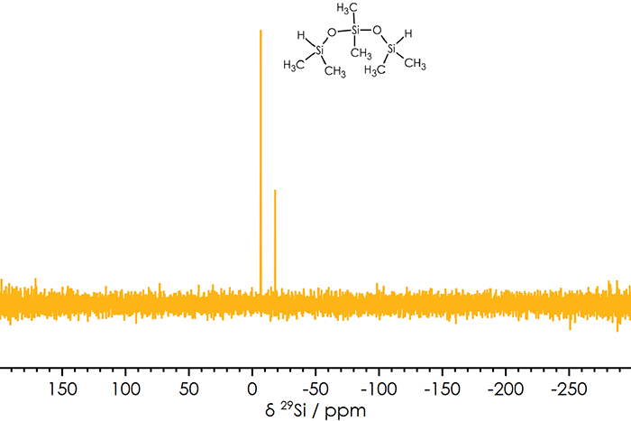
Figure 7a 29Si{1H} NMR spectrum of 1,1,3,3,5,5-hexamethyltrisiloxane.
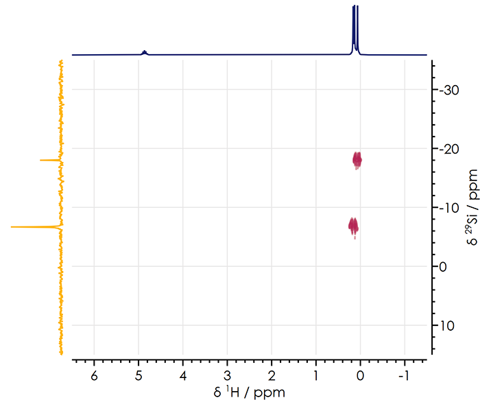
Figure 7b 1H-29Si HMBC NMR spectrum of 1,1,3,3,5,5-hexamethyltrisiloxane.
4one bond proton-silicon couplings can be obsereved in a HSQC spectrum
Cobalt-59 NMR Spectroscopy
Cobalt-59 (59Co) played an important role in the development of NMR spectroscopy, as the first nucleus for which the variation in chemical shift for different compounds was observed. Since many cobalt compounds are paramagnetic, 59Co NMR spectroscopy is generally limited to low-spin cobalt(I) and cobalt(III) complexes, which are observed over the largest chemical shift range of any nucleus, δCo −4,000 to +14,000 ppm 5, making 59Co an interesting nucleus for educational purposes. 59Co NMR spectroscopy also has applications for research and development in organometallic and coordination chemistry, along with studies of cobalt containing metalloenzymes. In this example (Figure 8), chemical shifts of the cobalt(III) complexes [Co(CN)6]3− and [Co(en)3]3+ (en = ethylenediamine) are observed to be separated by 7093 ppm.
![59Co NMR Spectra of [Co(en)3]3+ and [Co(CN)6]3− in aqueous solution.](/learning/uploads/inline-images/x-nuclei-part2-figure8-20260622125322.png)
Figure 8 59Co NMR Spectra of [Co(en)3]3+ and [Co(CN)6]3− in aqueous solution.
5This corresponds to 255 kHz on the X-Pulse 60, or 356 kHz on the X-Pulse 90; on high-field spectrometers the frequency range is so large (> 1 MHz) it’s rarely possible to acquire the entire range in a single spectrum
Platinum-195 NMR Spectroscopy
Platinum containing compounds have many applications, including as pharmaceuticals and catalysts, and 195Pt NMR spectroscopy is widely used for studies of these systems. 195Pt signals are observed over a wide range of 15,000 ppm (relative to Na2[PtCl6] in D2O at δPt 0 ppm), with the chemical shift strongly dependent on the oxidation state and coordination environment of the platinum centres.
For example, spectra of the half-sandwich platinum(IV) complex, [Pt(η5-C5H4CH3)(CH3)3] (Figure 9), shows a distinct signal in the 195Pt{1H} NMR spectrum at δPt −5240 ppm; with coupling to the nine-equivalent protons of the three methyl ligands clearly resolved in a proton-coupled 195Pt spectrum. While in the corresponding proton NMR spectrum, signals with distinct platinum-satellites from the 34% abundant 195Pt are observed.
![195Pt NMR spectra of [Pt(CH3)3(η5-C5H4CH3)], with and without 1H decoupling](/learning/uploads/inline-images/x-nuclei-part2-figure9a-20260622125819.png)
![1H NMR spectra [Pt(CH3)3(η5-C5H4CH3)], with and without 195Pt decoupling](/learning/uploads/inline-images/x-nuclei-part2-figure9b-20260622125839.png)
Figure 9 195Pt NMR spectra of [Pt(CH3)3(η5-C5H4CH3)], with and without 1H decoupling (above); and 1H NMR spectra [Pt(CH3)3(η5-C5H4CH3)], with and without 195Pt decoupling (below).
Summary
Broadband NMR spectroscopy allows for the observation of a wide range of chemical elements / nuclei, applicable to many different applications and markets. The Oxford Instruments X-Pulse 90/60 Spectrometers brings fully tunable broadband NMR to the benchtop.

