Using Confocal Microscopy to Pursue the Fundamental Stages of Developmental Biology
Author: Dr Claudia Florindo
Published: 01 Nov 2021 · Last updated: 18 Feb 2025
In this case study, we present an overview of scientific and technical challenges in thyroid hormone research. We introduce BC43, the new benchtop confocal from Andor Technology, and show how BC43 helps to overcome challenges in developmental biology research.
This article is divided into three sections:
Andor Benchtop Confocal Microscope - BC43;
Overview of the role of Thyroid hormone in metamorphosis;
Application case study: Research on molecular mechanisms of development and metamorphosis.
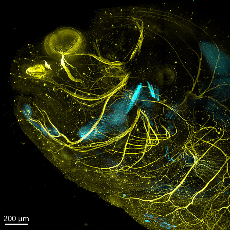
Figure 1 – Flat fish head imaged with BC43 using confocal imaging mode. Metamorphosing sole larvae were stained with acetylated tubulin to depict neuron (Yellow), and striated muscle using an anti-myosin heavy chain serum (cyan). This image illustrates the high detail and resolution of individual axons (yellow). Image was acquired with Andor Bench Top Confocal – BC43 using multiple tile acquisition, montage and a long working distance 40X objective. 48 tiles were acquired, had 175 slices, over a Z range of 330 µm, the resulting acquisition was deconvolved and stitched. Perfect stitching can be observed. Image was further rendered with Imaris (Image by Marco Campinho, CBMR Universidade do Algarve and Claudia Florindo, Andor Technology)
Introduction to Andor Benchtop Confocal - BC43
BC43 is Andor’s new plug and play high-speed confocal system and is a benchtop confocal imager that delivers remarkable imaging excellence at a unique price. With the BC43, the user can choose different imaging modalities according to the experimental goals. Imaging modalities include:
High quality confocal fluorescence imaging
Widefield epifluorescence imaging
Laser free imaging (Transmitted light)
BC43 is an ideal system for multiple life sciences applications. One such application is developmental biology. BC43 cuts through the imaging challenges of the Developmental biologist’s three-dimensional world.
Researchers can easily study development from the first rounds of cell division to the entire organism. It delivers fast, high-resolution imaging of developing model organisms. Furthermore, it images deeper than conventional fluorescence microscopes and images 10-times faster than traditional confocal systems.
Movie 1 - Live Imaging of Zebrafish Vascular development. Live image of transgenic zebrafish vascular development (shown in Cyan) combined with DPC (differential phase contrast) in grey. Imaging was performed from 28hpf until 36hpf. Image was acquired with using the confocal imaging modality of Andor confocal spinning disk – BC43. The imaging protocol had the time-lapse and montage activated. Nine tiles were acquired at each time point, for both the confocal and DPC channel, a range of 633 µm is covered. The resulting image was deconvolved and stitched using Imaris software. (Image by Marco Campinho Universidade do Algarve and Claudia Florindo, Andor Technology).
The main challenges encountered in developmental biology and how these are addressed by the Andor benchtop confocal BC43 are summarised in the following table:
Challenge
Solution
Thick specimens How to image deep inside thick samples
1. Optimized pinhole spacing and size allow high background rejection, delivering a high signal to noise for images over the 100(s) of micrometres range.
Live imaging How to perform live imaging without phototoxicity or photobleaching
1. Dual micro-lens multi-pinhole system withhighly efficient excitation filter and detectors allow sensitive imaging which results in minimal phototoxicity and photobleaching. 2. Live imaging of developing organisms can be accomplished for periods over 48h.
Large sample imaging Image through large samples Image multiple live samples Acquire significant amounts of data within a short, effective time frame.
1. Large Field of view (18.4 mm) allows increased productivity (less images needed to acquire the whole sample) 2. Integrated motorized stage and stitching lets you capture the complete development of the organism and stitch the data on the fly. The full image will be built as acquisition is proceeding. 3. Multiposition multipoint option allows acquisition of multiple organisms in a single experiment – increase productivity. 4. Multipoint confocal, delivers confocal images as fast as 44 fps, and 10X faster than regular point scanners. Increase productivity for both fixed and live imaging experiments.
Dr Marco Campinho (Group leader at the University of Algarve) is a developmental biologist whose research focuses on the molecular mechanisms of metamorphosis. Specifically, Dr Campinho aims to understand how the thyroid hormone controls this development process. In his research, he images large model organisms (zebra and flatfish).
We asked Dr Campinho to test the Andor benchtop confocal - BC43 to see how well it would work for these kind of developmental biology studies. After testing our system, Dr Campinho stated that:
“I have been using widefield, point scanners and light-sheet microscopes from the major brands for live imaging over the last ten years. Yesterday, it took me only 30 min to prepare eight embryos for live imaging over 24h. It was the fastest, easiest way that I have ever experienced in my career. I really love the BC43 system.”
Continue reading to get a grasp on current research in molecular mechanisms of metamorphosis and the technologies used to understand this fundamental developmental stage.
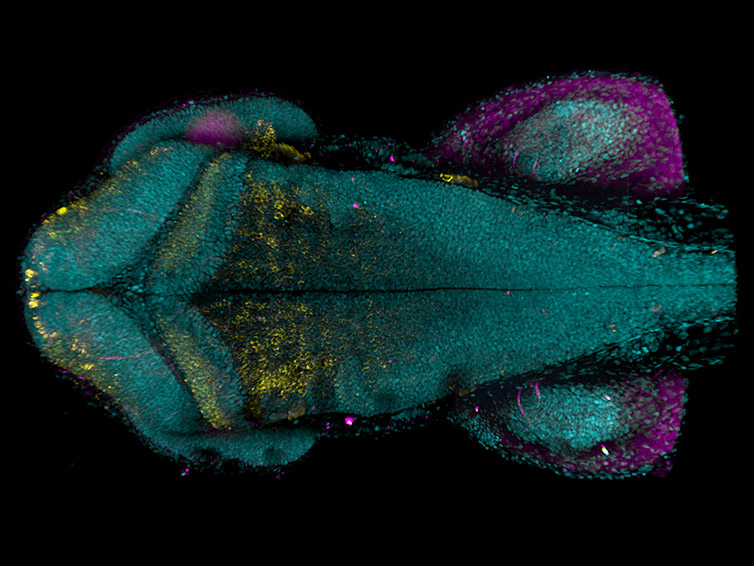
Figure 2 – Whole-body Zebrafish at climax of development. Maximum projection of zebrafish hindbrain at 48hpf after immuno-labelling neurons (anti-HuC/D serum, yellow), glia (anti-zrf serum, magenta). Nuclei were labelled with DAPI (cyan). Image was acquired with Andor benchtop confocal – BC43 using multiple tile acquisition and montage. 30 tiles were acquired to compose the image, each tile had 175 slices, over a 370 mm Z range. (Image by Marco Campinho Universidade do Algarve).
Overview of Thyroid Hormone Role in Metamorphosis
Hormones are essential molecules that regulate all multicellular organisms. Hormones can be described as “messenger molecules” produced and secreted by the endocrine system that control the homeostasis of the whole organism. The endocrine system comprises many glands; each produce specific hormones. The thyroid is one of the glands of the endocrine system.
The thyroid produces the T4 hormone (the precursor of the thyroid hormone). T4 will undergoes local activation giving rise to T3 (the active thyroid hormone). The thyroid hormone function in vertebrate physiology has importance across all life stages from embryonic development through to adult homeostasis.
Among the various developmental roles reliant on the thyroid hormone, human brain development stands out. Low levels of circulating thyroid hormones in pregnant women lead to severe neurological defects in new-borns. Several other diseases have also been associated with the de-regulation of the thyroid system, such as Graves disease, Hashimoto Thyroiditis, and more recently, roles in clinical depression are becoming clear.
The thyroid hormone is very important for vertebrate development and physiology; its developmental role was first established in the metamorphosis of Anuran amphibians (Allen, 1925). Further studies indicated that, as in anurans, T3 is essential for bony fish (teleosts) metamorphosis (Inui and Miwa, 1985). More recently, the Campinho laboratory proved that T3 is an essential regulator of embryonic development, whose role is conserved from fish to mammals (Campinho et al., 2014).
Modern research has established the thyroid hormone as a key regulator of development. Still, much more research needs to be undertaken to fully grasp the regulation and nuances involved in T3 developmental function. As T3 thyroid hormone has such a large impact on the overall development and homeostasis of vertebrate organisms, it is obvious that a better knowledge of T3 function as a whole will allow the development of therapeutic directed studies that will contribute to ease the burden associated with T3 related disorders.
Glossary
Allan-Herndon-Dudley syndrome
Allan-Herndon-Dudley syndrome is a brain development disorder, which results inmodest to acute intellectual disability and problems with movement. This condition is an X-linked disorder and is caused by a defect in a specific T3 hormone transporter (MCT8).
Anuran
Anuran is an order of amphibians characterised by having no tail in the adult stage. Examples of Anurans are frogs, toads, and tree frogs.
endocrine system
The endocrine system is composed by the glands that produce the hormones. The hormones are secreted to the bloodstream to act as messengers that ensure overall organism homeostasis and function.
hormones
Hormones are chemical elements that regulate physiological activities and maintain general organism homeostasis. At their core, hormones function as messengers. Hormones are secreted to the blood stream and will act to control and coordinate activities throughout the body.
Metamorphosis
Metamorphosis is a biological process in which an organism will suffer noticeable and sudden transformations in its body structure, encompassing development of immature organs to fully developed adult tissues. One example of metamorphosis is when the caterpillar becomes a butterfly.
Phenocopy
A phenotypic feature (or disease) expressed by an individual that is similar to the feature expressed by a particular genotype. Importantly, the individual who presents the feature is not a carrier of that genotype. The phenocopy occurrence can be employed in drug discovery developments — using the inhibition of a drug target and analysing the effects on the phenotype of interest
T3
T3 – triiodothyronine is the active form of the thyroid hormone
T4
T4 - thyroxine is the precursor of the Thyroid hormone (T3) that is produced in the Thyroid gland.
teleost
Teleost is the common aquatic species of fish. Around 96% of fish species known are teleosts. Teleost fish are characterised by the bone skeleton and the caudal fin which presents a symmetrical fork structure.
Thyroid
The thyroid is a gland localized in the neck. The thyroid produces hormones triiodothyronine (T3) and thyroxine (T4), whose function is to regulate the organism’s development and metabolism.
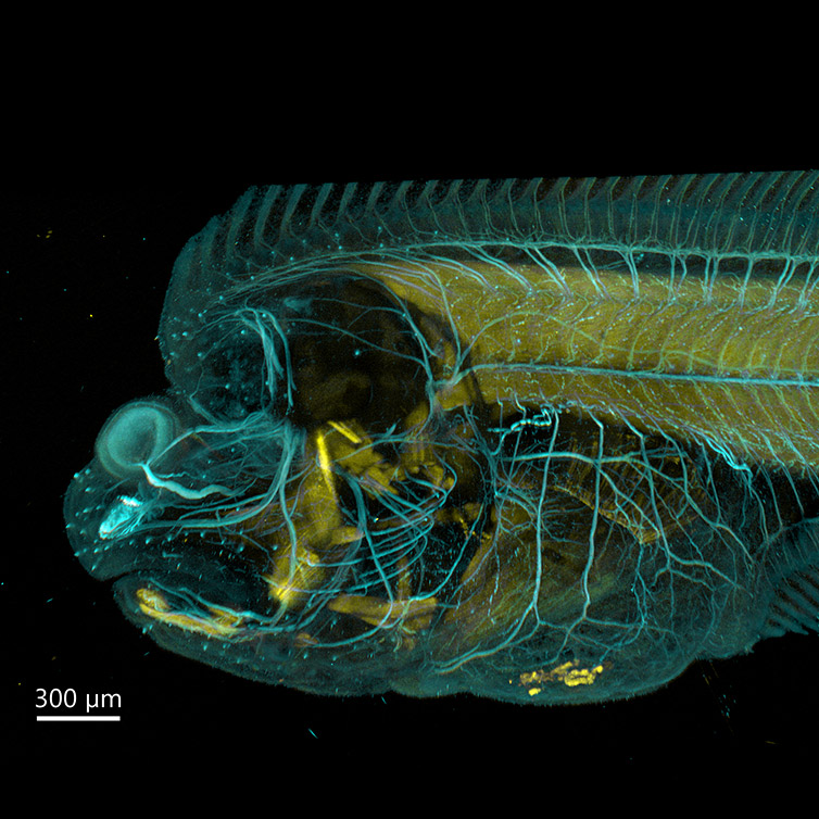
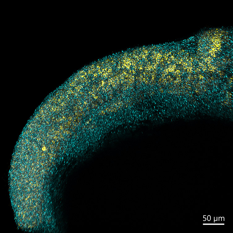
Figure 3 – Flat fish imaged with BC43. Flatfish at the beginning of climax stage of metamorphosis stained with acetylated tubulin for neuron labelling (cyan) and striated muscle myosin heavy chain (yellow). Image was acquired with Andor Bench Top Confocal – BC43 using multiple tile acquisition and montage. 6 tiles were acquired to compose the image each tile had 175 slices, over a Z range of 521 µm. Image was further rendered with Imaris. (Image by Marco Campinho Universidade do Algarve and Claudia Florindo, Andor Technology).
Application Case in Developmental Biology - Research on Molecular Mechanism of Development and Metamorphosis
The Campinho laboratory is interested in understanding the cellular and molecular basis of vertebrate development from embryonic development to metamorphosis. Given the widespread role of T3 in development (including embryonic development and metamorphosis), the laboratory has decided to focus the research on thyroid hormone molecular function. To pursue the research goals, the laboratory uses zebrafish and flatfish as model organisms.
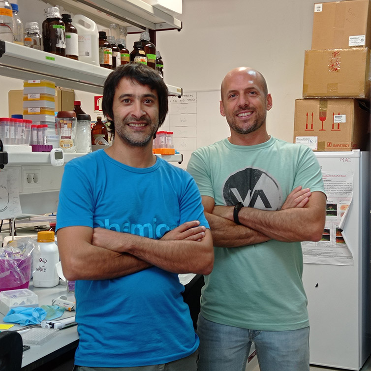
Figure 4 – Marco Campinho Group Leader (left) and Andre Andrade PhD candidate (right) in the lab.
Currently, the laboratory has two main ongoing research projects:
1) How does T3 hormone generate cell diversity during zebrafish neurodevelopment?
Understanding the molecular mechanisms of T3 in the neural development of zebrafish will drive knowledge on the overall molecular and cellular mechanisms of T3 signalling.
The Campinho laboratory has shown that maternal derived T3 is essential and is responsible for generating the necessary cell diversity and consequently promoting the development of a fully functioning central nervous system (Campinho et al 2014). Using zebrafish, the laboratory has developed the first vertebrate model for the Allan-Herndon-Dudley syndrome. Importantly, the fish model allows phenocopying (to mimicing) the neurological symptoms observed in human patients.
For the future, the laboratory aims to further develop the zebrafish loss-of-function mutants for T3 transporters and receptors; and expects that the knowledge gained from these studies will contribute to a deeper understanding of T3-related syndromes.
Figure 5 – Zebrafish hindbrain. Image shows double fluorescence in situ hybridization of a 24h zebrafish hindbrain. In Yellow – delta A from the notch signalling pathway and in cyan - Thyroid receptor (thraa). Image is a maximum intensity projection of 139 stacks covering a range of 187 mm depth. Image was acquired with Andor benchtop confocal - BC43. Image by Marco Campinho, Universidade do Algarve.
2) How does flatfish metamorphosis lead to the development of an asymmetric vertebrate?
The Campinho research group is also focused on understanding the basic mechanisms of metamorphosis.
Using the flatfish Solea senegalensis, the laboratory focused on understanding what mechanisms drive the asymmetric metamorphic development. The group was able to unravel the molecular and cellular mechanisms underlining the asymmetric metamorphic development of flatfish, where migration of one eye to the other side of the head results from asymmetric T3 signalling and ossification (Campinho et al., 2018).
Overall, the Campinho laboratory aims to address the cellular and genetic mechanisms that drive thyroid hormone regulation of vertebrate development. Specifically, the laboratory aims at deciphering:
The relevance of T3 signalling and its translation to human diseases.
The fundamental mechanisms of morphogenesis.
Not surprisingly, the laboratory uses a variety of molecular and imaging techniques to achieve the goals including: 1) zebrafish transgenic and CRISPR/Cas9 genetic models, 2) pharmacological approaches, 3) OMICs and advanced microscopy techniques.
The group has been using long-term (40 hr) live imaging of developing zebrafish embryos as well as developing clearing techniques to allow whole-mount high-resolution imaging of metamorphic flatfish larvae using the BC43 imaging system. According to Dr Campinho
“Microscopy wise we have to supress several complex technical hurdles. Namely in metamorphosis given the large size of the samples to be imaged (1-1.7 mm thickness). “
We asked Dr Campinho to comment further on testing our new Andor benchtop confocal - BC43 for his research in thyroid hormone regulation:
“Andor spinning disk technology provides the necessary microscopy technology to allow surpassing of the technical challenges at hand. “
Thank you very much for your feedback Dr. Campinho. We look forward to seeing more of your fantastic imaging and interesting scientific research.
Bibliography
Allen, BM. The effects of extirpation of the thyroid and pituitary glands upon the limb development of Anurans. Journal of Experimental Zoology 1925; 42:13-30.
Campinho, MA, Saraiva, J, Florindo, C and Power, DM. Maternal thyroid hormones are essential for neural development in zebrafish. Mol Endocrinol 2014; 28:1136-1144.
Campinho, MA, Silva, N, Martins, GG, Anjos, L, Florindo, C, Roman-Padilla, J, Garcia-Cegarra, A, Louro, B, Manchado, M and Power, DM. A thyroid hormone regulated asymmetric responsive centre is correlated with eye migration during flatfish metamorphosis. Scientific Reports 2018; 8:12267.
Inui, Y and Miwa, S. Thyroid hormone induces metamorphosis of flounder larvae. Gen Comp Endocr 1985; 60:450-454.