Aim
Detection of contamination in the constituent materials used to make batteries is critical for performance, safety and economic reasons. Impurities, such as Ti, S and Cu particles in raw materials can have significant impacts on electrochemical performance and material stability and can even cause internal shorts and subsequent severe safety issues. Finding and identifying contamination in a timely manner allows the source to be determined and corrective actions implemented with the minimum of downtime for production.
Solution
Initial Sample Investigation
In many cases, an initial, manual review of a sample provides the necessary information to understand if key contaminants are present. Given the size and nature of critical contaminants, an analytically equipped scanning electron microscope (SEM) is an ideal tool for the job. Traditionally, this approach has been laborious owing to the linear nature of the workflow which required sequential electron imaging of the sample, investigation of composition with Energy Dispersive X-ray Spectrometry (EDS) followed by moving of the sample to a new site and imaging again. This makes the approach impractical for routine use.
The solution to this is to rapidly review the sample with a SEM equipped with Unity, as the combined Backscattered Electron (BSE) and very high count-rate X-ray (BEX) imaging method means that potential contaminants can be reliably picked out and identified by both morphology and composition even when they are very small (i.e. <1 µm). The BEX approach can be utilised in real time, as the very high throughput means that detailed combined imaging can be performed even whilst the sample is in motion during navigation around the sample. For potentially contaminated battery powder samples, this means that important decisions can be made on whether production should proceed or on where corrective actions needs to be taken very rapidly.
The distribution of particles of multiple types within the electrode is seen immediately via BEX imaging, as both topographic and compositional information are displayed simultaneously, thereby reducing time-to-report significantly (Figure 1).
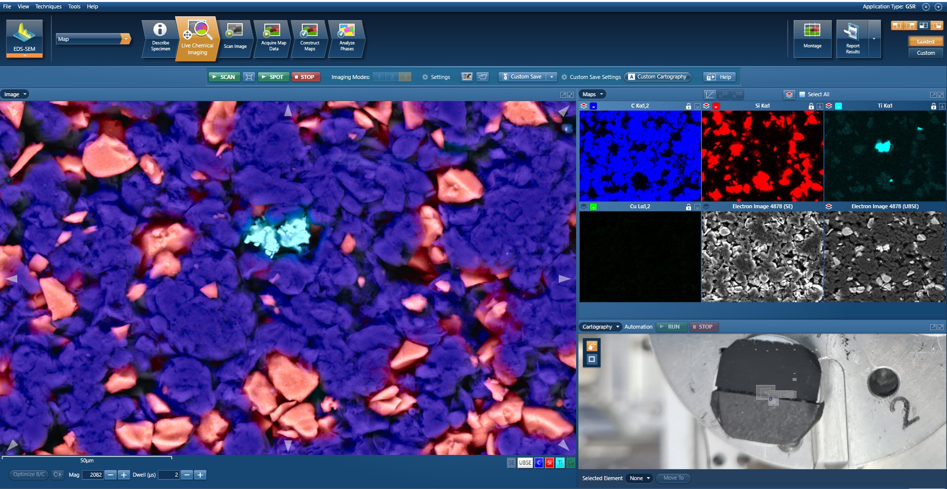
Figure 1. BEX Imaging of a battery anode. The C and Si parts are easily imaged as is a Ti contaminant
Challenging Feature Positioning
The under-polepiece design of Unity means that BEX imaging is performed from a top-down perspective. This means that even when particles or features of interest are present in topographic lows within samples, they are still accurately imaged for their composition. This is a great advantage when searching for contamination, as grains of different morphology to those that make up the majority of the sample will frequently fall into topographic lows – and can be almost impossible to identify by other means (e.g. the Ti in Figure 1).
High Quality Imaging Output
Once a particular contaminant has been identified, the next step is to communicate its presence to others so that the appropriate remedial action can be taken. This is best done with a high-quality BEX image that clearly shows the topographic and compositional properties of the contaminant. High-quality images of this type can be easily produced with Unity – with large quantities of compositional X-ray data combined with BSE imaging to produce images that clearly show exactly what contamination is present and in what form. Examples of such images are shown in Figure 2 and Figure 3; each was produced in a matter of a few seconds.
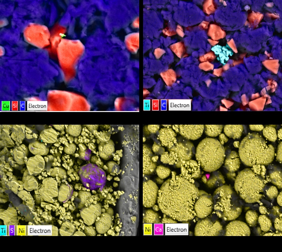
Figure 2. Rapid, high-resolution BEX imaging with Unity. Publication quality BEX images show multiple contamination types in anode (top) and cathode (bottom) samples. All images were acquired in <5s. Image (a) shows Cu contamination smaller than 1 µm detected in an anode sample as well as (b) larger clusters of Ti – which are clearly compositionally imaged, even when in a hole. Image (c) shows precipitated sulphur contamination coating a larger NCM particle as well as very small titanium particles in the upper and lower left parts of the image. Image (d) shows a micron sized grain of calcium sulphate compositionally imaged even though it is in a valley between particles.
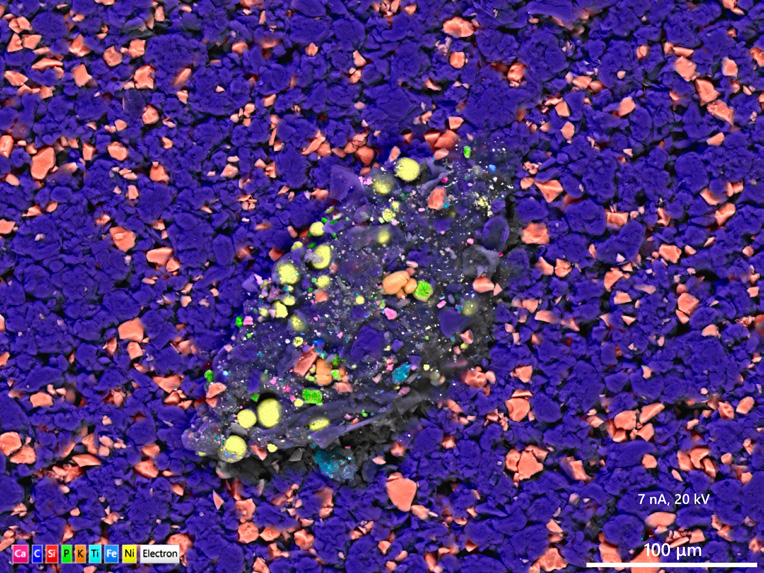
Figure 3. BEX high resolution custom save of an unexpected contaminant powder grain in an anode sample.
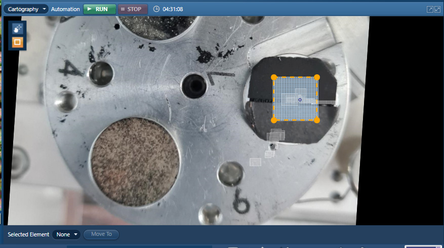
Figure 4. Setup of a BEX run with Cartography Mode. The area to be analysed (outlined in dashed orange) was dragged out over the registered image with the run started by hitting the “run” button.
Large Area BEX Imaging
Many contaminants will be (hopefully) rare within the sample and, therefore, may not be immediately visible in any particular field of view studied with BEX. This makes it very important that either a large proportion or all of a sample is analysed to determine if contaminants are present. In many cases, the process of doing this will reveal not only known contaminants but also previously undetected ones.
It is a quick and easy process to move from single field BEX imaging to multi-field analysis of a complete sample. By making use of cartography mode, a large area can be set up for analysis with a single mouse drag across a registered image. This is performed by simply dragging the mouse cursor over the area of interest on the screen (Figure 4). The BEX analysis across the entire defined area then runs immediately with simultaneous BSE and X-ray data collected for every pixel. This has a significant advantage over traditional mapping of samples as, due to the very high X-ray collection rate, the X-ray data can be acquired in the same time as the BSE image. This means that the acquisition of BSE and compositional data takes place in a single step as opposed to sequentially – with a consequential and very significant throughput improvement achieved.
Results
BEX imaging with Unity provides both topographic and compositional information simultaneously, allowing for rapid, intuitive electrode structure characterisation. Quick and reliable analysis of samples of this type has shown the presence of impurities, such as Ti, Cu, S, Cu, & Al, even down to a sub-micron size. Unity provides both exceptionally powerful BEX imaging for locating contaminants in both single fields of view and across large areas (Figure 5) as well as fast, publication quality compositional imaging output, even under challenging circumstances – such as the analysis of particles in valleys.
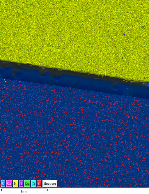
Figure 5. Large area BEX imaging acquired with Unity in AZtecLive's Cartography Mode. The area consists of 608 fields and was collected in 1 hr 33 min.

