Raman microscopy in core facilities: Key benefits and applications
Core facilities have become central research infrastructures across academia and industry. Rather than functioning as simple equipment rooms, they increasingly act as interdisciplinary technology hubs that support materials science, semiconductor research, life science, battery development, environmental analysis, and pharmaceutical applications within shared environments.
As the diversity of users continues to grow, expectations around automation, reproducibility, usability, and multimodal data integration are also increasing. In this context, confocal and correlative Raman imaging platforms play a particularly important role. They combine chemically specific imaging with high spatial resolution, while remaining flexible enough to support different workflows and multiple analytical techniques on a single platform.
In the following interview, Sabrina Hakelberg, Product Manager for Oxford Instruments' Raman product lines, discusses how modern Raman imaging systems are evolving to meet both the scientific and operational requirements of today's core facilities.

Sabrina Hakelberg, Product Manager for Raman microscopes at Oxford Instruments
Why are institutes investing in centralised imaging and spectroscopy platforms?
Sabrina: Centralised platforms allow institutes to share highly advanced technologies across multiple departments. This helps avoid redundant investments and maximises overall instrument utilisation, creating both economic and scientific benefits. At the same time, research has become increasingly interdisciplinary. A single facility may support physicists, chemists, biologists, engineers, and industrial collaborators.
As a result, core facilities are evolving from equipment-centred spaces into expert-driven research infrastructures that provide consultation, workflow development, training, and standardised analytical services. This shift directly influences what core facility managers expect from their instruments. Advanced imaging and spectroscopy systems must deliver high analytical performance, while also operating reliably in multi-user environments with very different levels of user expertise.
Confocal Raman imaging platforms are very well suited to shared environments, because a single system can support exploratory research, routine characterisation, and correlative workflows across a wide range of scientific disciplines.
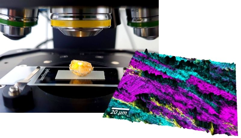
Human gallstone under an Oxford Instruments Raman microscope. In the topographic Raman image, two cholesterol derivatives are coloured in cyan and pink. Sample courtesy of Miriam Böhmler, Oxford Instruments.
What makes Raman imaging microscopy attractive for core facilities?
Sabrina: Raman imaging provides chemical analysis with micrometre-scale spatial resolution. It is label-free and non-destructive, meaning the sample remains unchanged during measurement. This is especially valuable in shared infrastructures, where one platform often needs to support many different application areas. Typical examples include studies of semiconductors and advanced materials, batteries and energy materials, polymers and composites, particles and contaminations, pharmaceuticals, environmental samples, and cells and tissues.
Another important advantage is that Raman data integrates naturally into microscopy workflows. Users work with chemical maps, overlays, regions of interest, and correlative imaging approaches that are already familiar from optical microscopy.
For core facilities, the broad applicability of Raman imaging increases instrument utilisation and supports interdisciplinary collaboration across departments and external partners.
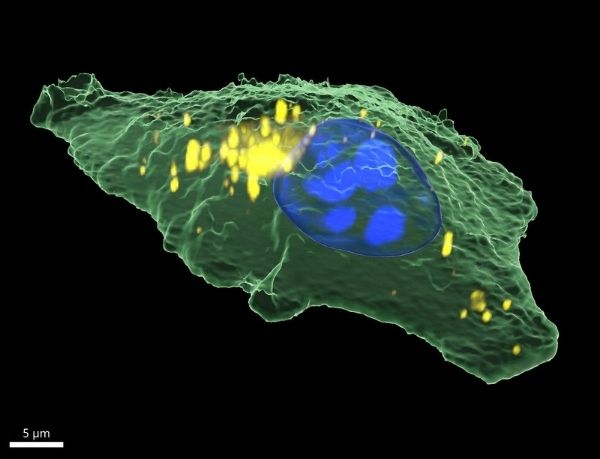
3D Raman image of a human epithelial cancer cell showing cytoplasm (green), nucleus (blue), and lipids (yellow). Dimensions: 40 x 47 x 9 µm³. Sample courtesy of Dr. Irina Estrela-Lopis and Tom Venus, Institute of Medical Physics and Biophysics, Leipzig University, Germany.
What is the main challenge in multi-user environments, and how do modern Raman platforms address it?
Sabrina: The main challenge in shared environments is delivering high analytical quality and flexibility, independent of individual user expertise. Historically, Raman microscopy was often seen as highly expert-driven. Measurements can be sensitive to alignment, spectral configuration, calibration, and acquisition parameters. In core facilities, however, systems must perform reproducibly across many users and application types.
Modern Raman platforms address this challenge by making automation a central design principle. They increasingly integrate automated alignment and calibration, guided acquisition workflows, predefined measurement templates, automated spectrometer configuration, and standardised reporting procedures.
For example, modern spectrometer concepts such as Hexalight allow automated switching between spectral configurations without manual realignment. Automated optical alignment and calibration routines further reduce setup complexity and improve reproducibility.
The goal is not simply convenience. It is operational robustness. Automation features allow facilities to generate reliable and reproducible Raman datasets regardless of operator experience.
How important are correlative imaging workflows in core facilities?
Sabrina: Correlative imaging has become one of the strongest strategic drivers for advanced microscopy platforms. In many applications, a single measured parameter is no longer sufficient. Researchers benefit from combining structural, chemical, and functional information within one coordinated workflow.
Raman imaging and spectroscopy play a unique role in this context because they add direct chemical specificity to well-established imaging techniques. For example, integrated systems enable correlative Raman, SEM (Scanning Electron Microscopy) and EDS (Energy Dispersive X-ray Spectroscopy) for defect analysis and contamination studies. Raman and fluorescence microscopy are popular in biological research, and Raman combined with nonlinear SHG (Second Harmonic Generation) is used for analysing collagen fibres or for interface studies. Topography-guided imaging enables Raman mapping of rough and structured surfaces, and polarisation-resolved Raman and white-light imaging is powerful for analysing anisotropic materials and tissues.
In materials science, these combined approaches are used to study crystal orientation, strain domains, composite structures, and semiconductor defects. In life science, similar concepts are applied to collagen alignment, tissue organisation, and structural heterogeneity in biological samples.
From a core facility perspective, correlative workflows are particularly valuable because they increase information density while reducing the need for redundant measurements across multiple instruments.
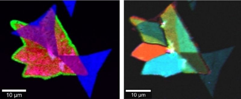
Raman and SHG images of a 2D material heterostructure. The Raman image (left) identifies areas of MoS₂ (blue) and WS₂ (red) crystals, and overlapping areas (purple), as well as edge effects (green). SHG (right) recorded at three different polarisation angles (red, green, blue) shows grain boundaries and grain orientations.
How can Raman imaging serve a broad range of research groups in core facilities?
Sabrina: The overlap between the requirements of different research groups is often greater than expected. Many analytical workflows are fundamentally similar, even when the samples and scientific questions differ, and can thus serve diverse research groups.
Raman-assisted particle analysis is a good example. In materials science and industrial research, users investigate contamination particles, wear debris, polymer fragments, or process residues. In environmental and life science research, very similar workflows are applied to microplastics or particulate structures in complex matrices. In both cases, the analytical process follows the same basic steps: detect particles, classify morphology, identify chemical composition with Raman, and generate statistically robust datasets.
Other examples are polarisation-resolved and nonlinear optical imaging techniques, such as SHG and THG (Third Harmonic Generation), used alongside Raman imaging. Materials researchers apply these methods to study anisotropy, crystal orientation, or stress-related ordering. Life science researchers use comparable approaches to investigate tissue organisation, collagen alignment, and fibre orientation.
The underlying physical principles of these analytical techniques are therefore often shared across disciplines, even when the specific research goals differ.
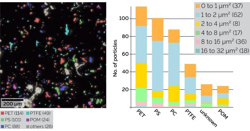
Raman-based microplastic particle analysis: Dark-field image with microplastic particles colour-coded according to their Raman spectra (left), and particle size distribution (right).
What do core facilities expect from technology partners?
Sabrina: Core facilities increasingly look for long-term scientific and operational partnerships, rather than purely transactional vendor relationships. This typically includes application support, workflow development, training programmes, support for grant proposals, responsive technical service, and assistance with method standardisation.
Facilities also place growing emphasis on scalability and long-term sustainability. Platforms are expected to evolve with changing research priorities, increasing user numbers, and expanding analytical requirements.
As a result, purchasing decisions are no longer based solely on peak instrument specifications. Decision makers increasingly evaluate factors such as operational reliability, ease of onboarding, interdisciplinary applicability, automation capabilities, and long-term adaptability.
Systems that succeed in core facility environments are therefore typically designed as scalable platform technologies, rather than narrowly optimised single-purpose instruments.
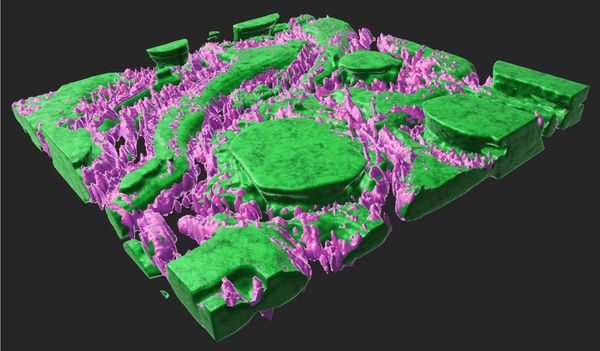
3D confocal THG imaging reveals the distribution of adipocyte cells in a subcutaneous tissue sample. Cells are shown in green and cell membranes in purple. Dimensions: 65 x 65 x 7 µm³. Sample courtesy of Prof. Jiang Chunhuan, Changchun Institute of Applied Chemistry, CIAC CAS.
How will Raman imaging platforms evolve?
Sabrina: Future development will strongly focus on workflow integration, automation, and data infrastructure. Core facilities increasingly require higher throughput, greater reproducibility, easier onboarding for non-expert users, and seamless integration into multimodal imaging environments.
This will drive further advances in areas such as AI-assisted analysis, more automated analysis workflows, intelligent workflow guidance, and integrated data management aligned with FAIR principles.
At the same time, correlative imaging will continue to expand. Raman microscopy is increasingly positioned not as a standalone technique, but as a chemical imaging layer integrated into broader microscopy ecosystems.
Ultimately, the most successful platforms will combine high analytical performance with operational simplicity, scalability, and interdisciplinary flexibility. This allows core facilities to support both scientific innovation and sustainable shared infrastructure models over the long term.
Conclusion: Raman microscopy in core facilities
Raman imaging microscopy has evolved from a specialised analytical technique into a versatile platform technology capable of supporting the complex requirements of modern core facilities.
By combining chemical specificity, confocal imaging, correlative workflows, automation, and scalable data handling, modern Raman platforms enable shared infrastructures to deliver reproducible, high-value analytical capabilities across disciplines.
For research institutions, the strategic relevance is clear: versatile imaging platforms are no longer just instruments. They are foundational technologies for collaborative and interdisciplinary research environments.
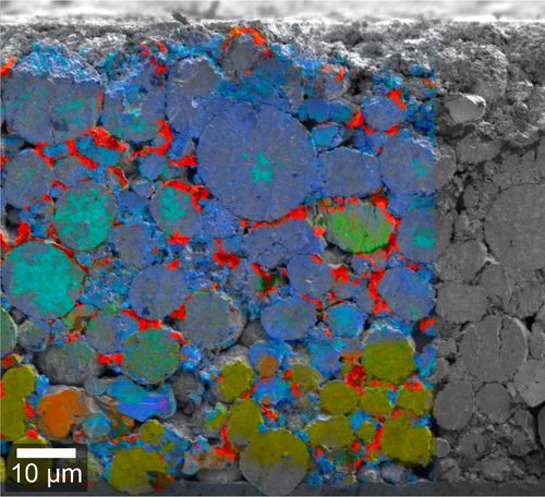
Overlay of Raman and secondary electron (SE) images of a polished lithium-ion battery cathode at 50% SOC, revealing LNO and NMC particle morphology and charge state heterogeneities. Sample courtesy of IMFAA Aalen, Germany

