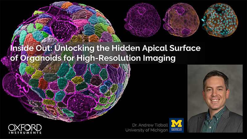
Organoid imaging requires microscopy techniques that can capture complex 3D structures with sufficient depth,
resolution, and throughput. Spinning disc confocal systems such as BC43 help overcome common organoid imaging
bottlenecks by combining optical sectioning, rapid acquisition, and low phototoxicity. Together with Imaris image
analysis software, BC43 supports a streamlined workflow from image acquisition to quantitative 3D analysis.
Introduction
Organoids have become indispensable for modern life science research. Beyond the limits of traditional 2D cell
cultures, they mimic key organ structures and functions and provide physiologically relevant 3D models to study
development, disease mechanisms, and therapeutic response. They are central to investigating the complex interplay of
different cell types and structures in biomedical research such as neurology, gastroenterology, and oncology, and play
a critical role in drug screening and early-stage clinical trials.
Yet, the structural complexity that makes organoids such valuable research models also introduce significant imaging
challenges. Organoids are typically thick, multicellular systems with internal luminal spaces and delicate structures.
Traditional microscopy often reaches its limits in capturing organoids in full detail, so more advanced solutions are
needed to provide the required detail, depth, and efficiency.
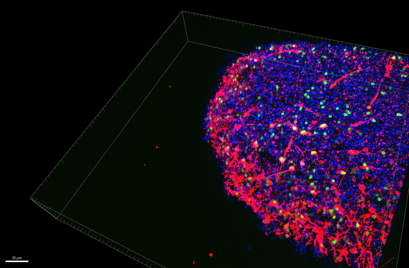
Human brain organoid. Scale bar 50 µm. Image credit: Yu Liu, University of Michigan
Common Challenges in Organoid Imaging
Organoids offer exceptional biological insight, but their size and complexity introduce significant hurdles for
traditional imaging workflows. Researchers commonly face the following challenges:
- Achieving depth while preserving structure: Organoids are multilayered structures that increase
light scattering and out-of-focus blur during imaging. Confocal microscopy enables optical sectioning to block
out-of-focus light and improve depth resolution compared to widefield imaging, even in living samples. However,
imaging deep inside thick samples often remains challenging. Physical sectioning can overcome this limitation, yet
slicing disrupts the 3D architecture, adds sample preparation complexity, and is incompatible with live studies.
- Capturing fine detail at realistic throughput: Point-scanning confocal imaging delivers
high-quality optical sectioning but can quickly become a bottleneck when imaging multiple positions or large
z-stacks. Modern organoid studies often require dozens of samples per condition to achieve statistical confidence,
which results in gigabyte- to terabyte-scale datasets. At this level, acquisition can take hours or days, delaying
analysis and limiting overall productivity. Multi-point spinning disc confocal systems help drastically shorten
imaging time.
- Minimising phototoxicity while maintaining resolution: Extended imaging sessions such as z-stacks
or time-lapse recordings across multiple channels increase the risk of photobleaching and phototoxicity, especially
in live experiments. In complex 3D systems, it is therefore often a challenge to balance illumination intensity and
exposure time while achieving sufficient signal strength.
These limitations often force researchers to compromise: Slice samples, reduce image volume, or limit replicate
numbers at the expense of data quality and significance or biological context.
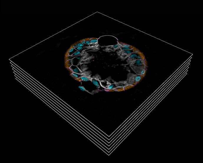
Stacked images of an LPA Colonoid. Image credit: Dr Andrew Tidball, University of Michigan
BC43 for High-resolution Organoid Imaging
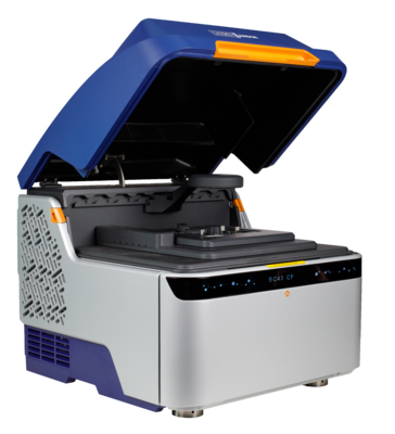
BC43 was designed to address the bottlenecks of organoid imaging through a combination of
spinning-disc confocal technology, high-sensitivity detection and ease of use, all in a compact and affordable
benchtop platform.
Key benefits for organoid imaging
- Fast optical sectioning at high spatial resolution: Capture complete high-resolution 3D stacks up
to 10x faster than in point-scanning confocal systems for efficient large-volume imaging in both live and fixed
samples
- Dual-microlens spinning disc technology for rapid, multi-point confocal imaging and minimal
phototoxicity
- High-speed sCMOS detection for sensitive imaging at short exposure times
- Scalable imaging with automated tile scanning: Move beyond single organoid fields and scale your
imaging pipeline
- Motorised XYZ stage with high precision for accurate positioning and mosaic acquisition
- Integrated stitching software for large-area coverage across wells and entire organoid-on-chip systems.
- Low phototoxicity and highly sensitive detection: Protect live samples during extended
experiments with optimised illumination and sensitive image acquisition
- Patented Borealis™ beam-shaping technology for uniform illumination across the entire field
of view
- High Sensitvity Andor sCMOS camera ensures excellent signal quality, even under low-light
conditions
- Live and long-term experiment compatibility: Monitor dynamic processes in organoids for hours or
days under stable environmental conditions
- Optional environmental chamber for precise control of temperature, CO2, and humidity during the experiment
- Laboratory-ready footprint and ease-of-use: Perform your imaging experiments directly on the
bench, with no darkroom requirement. BC43’s intuitive, user-friendly software makes advanced 3D workflows accessible
to experienced researchers and imaging newcomers.
Research Example: Whole-Chip Organoid Imaging with BC43
Researchers at the National University of Singapore used BC43 to image an entire organoid chip, which allowed them to
achieve results around 100 times faster than when using a conventional laser scanning confocal system, while
maintaining equivalent or superior signal-to-noise ratios. Complete datasets were stitched and rendered for
interpretation in Imaris within the same integrated pipeline. This approach streamlined an otherwise multi-hour
process into a routine workflow.
Organoid cultures grown on JeWell chip. Credit: Dr Anne Beghin, Mechanobiology Institute,
National University of Singapore
3D Image Analysis with Imaris
Acquiring high-quality images is only one part of an organoid imaging workflow. These workflows often generate
massive 3D datasets that quickly become unmanageable without the right tools. Extracting quantitative insight such as
lumen size, spatial organisation, or marker distribution requires advanced software that can efficiently process,
analyse, and visualise complex volumes.
This is why BC43 is paired with Imaris, Oxford Instruments’ trusted 3D/4D image analysis platform. Together, they form an integrated
pipeline from acquisition to interpretation.
Imaris capabilities for Organoid Research
- Real-time exploration of large datasets: Smooth handling of gigabyte-to-terabyte image volumes
with interactive 3D/4D navigation.
- Automated segmentation and surface rendering: Accurately identify organoid outlines and
substructures.
- AI-driven classification tools: Trainable algorithms to phenotype and categorise structures with
confidence.
- Batch workflows for reproducibility: Process multiple datasets consistently for high-throughput
imaging studies.
The result is an end-to-end solution, where powerful imaging and analysis work seamlessly together to turn detailed
imaging data into meaningful biological findings.
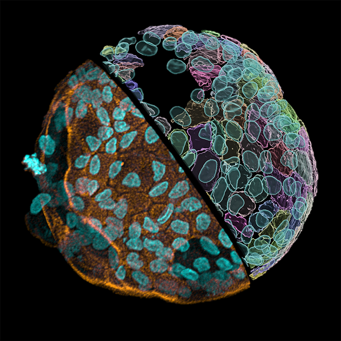
LPA Colonoid, imaged with BC43 and analysed with Imaris. Image credit: Dr Andrew Tidball, University of
Michigan
Application Spotlight: Studying Apical Polarity Cues in Neural Tube Organoids
BC43 supports organoid imaging across applications, from routine phenotyping to advanced studies of tissue
organisation and developmental biology. In a recent webinar, Dr Andrew Tidball from the University of Michigan Medical
School highlighted its use in neural development research. Dr Tidball and his team developed an organoid model to
explore mechanisms of apicobasal polarity during early neural tube formation, a process critical for proper neural
tube closure and central nervous system development.
In their work, the team generated iPSC-derived neural tube organoids and discovered a method to invert their
polarity, creating apical-out organoids. This finding enabled a detailed examination of polarity cues and led to the
identification of lysophosphatic acid (LPA) and its associated signalling pathway as key regulators of apical polarity
across multiple tissue types.
To analyse these polarity changes, the researchers needed high-resolution 3D imaging of the organoids in their intact
form. BC43 enabled rapid imaging of whole, immunostained organoids, providing fast overview scans at 20x magnification
and fine structural detail at 63x to visualise features such as apical junction proteins, actin belts, and cilia
markers. This approach allowed in-depth analysis of entire organoids without the distortions and time costs associated
with physical sectioning.
Watch the webinar to learn more about BC43 for organoid imaging and
Dr Andrew Tidball’s research on apical polarity mechanisms in neural tube organoids.