Tags: Thin Films and Coatings Nanoindentation, Mechanical Microscopy, Correlative Mechanical Microscopy
Mechanical microscopy of nuclear materials, such as hydrides at interfaces, is crucial for understanding the mechanical behavior of nuclear fuel cladding alloys. Hydrides in zirconium alloys can cause embrittlement and cracking, which can lead to fuel rod failures in nuclear reactors. Nanoindentation mapping is a valuable technique that provides information about the hardness and elastic modulus of the material at different points, including at the interface between the liner and substrate. This technique can be used to identify the presence and distribution of hydrides, as well as their effect on the mechanical properties of the material. In this application note, neutron radiography and mechanical microscopy were applied to investigate hydride behavior and distributions in samples with and without liners. This can help to improve the understanding and design of nuclear fuel cladding materials, ultimately contributing to the safety and reliability of nuclear reactors.
Introduction
Zirconium (Zr) alloys are commonly used for fuel cladding and structural components in nuclear reactors due to their mechanical properties and low neutron cross section [1]. Zircaloy-2 is a popular Zr alloy used as fuel claddings in boiling water reactors, some of which are produced with an inner liner to better cope with problems related to pellet-clad interaction and fission-gas corrosion. Despite the liners providing a major improvement in fuel reliability and operational flexibility, an increased tendency of failed fuel rods to exhibit post-failure degradation in the form of longer cracks has been observed due to the precipitation of hydrides in the cladding [2], which can occur in remote locations from the initial perforation of the fuel rod.
Hydride precipitation, growth, and orientation in nuclear cladding material is of high importance for the integrity of spent nuclear fuel and has been extensively studied in zirconium alloys [3]. Regardless of their pellet-clad interaction resistance, liners have been reported to act as a preferential nucleation site for hydrides.
High-resolution neutron radiography at the SINQ neutron spallation source at Paul Scherrer Institut was used to evaluate the hydrogen distribution present in the samples prior to controlled cooling. Nanoindentation mapping was used to characterize the phases present in the material and to reveal the hardness variations at the interfacial region. The influence of the different cooling rates on the hydrogen migration and precipitation was analyzed with respect to the hydride precipitation inside the liner and at the inner liner/substrate interface of the respective Zircaloy-2 samples.
In this application note, we investigate the capabilities of Mechanical Microscopy in conjunction with neutron radiography to assess the properties and distributions of hydrides in Zircaloy-2 cladding tubes with and without liners. Neutron radiography allows volumetric quantification of hydrogen content, while nanoindentation mapping allows quantification of area fractions of phases with different mechanical properties. The combination of these two techniques allows the measurement of mechanical behavior vs local hydrogen content. Here we explore the ability of these techniques to characterize local hydrogen concentration and mechanical effects.
Materials and Methods
Sample preparation
Two different types of commercial Zircaloy-2 cladding tubes, supplied by Framatome (LTP2 - unlined and LTP – with an inner liner) were prepared for this study. Tubular samples approximately 15 cm in length were enriched to hydrogen concentrations of 200 wppm. This was performed via diffusion of hydrogen into the sample material at high temperature in a Sievert-type apparatus, following a similar process to Fagnoni et al. [4]. The procedure consists of evacuating the chamber containing the sample to 10-7 bar, then exposing the sample at 400 °C to an atmosphere of 3–7 mbar of pure hydrogen. The hydrogen absorption by the sample was then monitored via the decrease in hydrogen pressure measured in the chamber.
Following hydrogenation, the specimens were subjected to a homogenization heat treatment at a temperature of 400 °C for 24 h in a furnace with a protective Ar atmosphere to create a uniform distribution of hydrogen throughout the specimens. Hydrogen contents were measured using hot gas extraction.
To investigate the hydrogen diffusion within the samples and hydride precipitation, selected rings were subjected to a second heat treatment consisting of 8 hours holding temperature at 400 °C. After this holding time, samples were directly exposed to different cooling rates: controlled furnace cooling at 3 and 30 °C/h and water quenching. Separate samples from several of these conditions, as well as the as received condition without hydrogen, were prepared for neutron radiograph and metallurgically prepared for microscopy and nanoindentation mapping. Results from quenched samples and samples cooled at 3 °C/h are discussed elsewhere [5].
Neutron radiography
The hydrides distribution within the samples before and after heat treatment was assessed by neutron radiography at the Swiss spallation neutron source, SINQ, at the POLDI beam line [6]. The neutron microscope used in this experiment is equipped with a 157Gd2O2S:Tb scintillator of 3.5 μm thickness and a CCD camera composed of 2048×2048 pixels. The nominal pixel size of the resulting radiographs is equal to 2.7 μm [7]. Both experimental procedure and post-processing follows the same methodology as Gong et al. [8].
Nanoindentation
Nanoindentation mapping was performed using an i04 Femto-Indenter with a diamond Berkovich indenter. A high-speed nanoindentation technique was employed to map local mechanical properties using continuous stiffness measurement (CSM) indentation testing [9] in displacement control. Each indentation was conducted in ≈1 s with an oscillation frequency of 140 Hz and an amplitude which was linearly increased with increasing depth from 1 to 2.5 nm. The total time for each indentation including repositioning was ≈2.5 s. Each indentation was performed to a specified depth of 190 nm, so that a spacing of 2 μm between indentations could be used while still ensuring an indentation depth/spacing ratio of ≈10 to avoid any significant interaction between neighboring indents [10]. Hardness, H, and reduced modulus, E, were measured as a function of depth for each location, and representative values for each parameter were taken by averaging values from depths >120 nm to minimize the influence of indentation size effects.
Electron micrographs were taken of each test region with a Zeiss EVO 25 scanning electron microscope (SEM) at 10 kV using a HD Backscattered Electron Detector (BSE).
Results and Discussion
Hydrogen distribution in cladding tubes by neutron imaging
High-resolution neutron images of unlined LTP2 and Fe-containing liner LTP, charged with a target concentration of 200 wppm in Hydrogen concentration, are shown in Figure 1. Radial neutron-transmission profiles were obtained from the high-resolution neutron images by circumferential integration over an angle of 30°. To quantify the hydrogen concentrations in all the measured samples, a calibration curve was used. All graphs display the neutron transmission on the ordinate axis from the evaluated neutron image area and its corresponding hydrogen concentration is plotted on the abscissa. The first 30-80 µm drop in neutron transmission at the sample edges is a consequence of neutron refraction from the sample surfaces. This edge effect is particularly relevant in the samples with liners, as it extends into a considerable fraction of the liner and can affect the calculation of the hydrogen content.
As shown in Figure 1, no significant changes are observed in the hydrogen content in the radial direction for the LTP2 clad tubes without liner. Transmission fluctuations corresponding to hydride clusters are clearly observable. The relatively good contrast means that the hydride clusters have a considerable depth within the sample, i.e. along the neutron traveling path, and the fluctuations become more visible in the slowly cooled sample. Hydrides tended to be longer and thicker when slower cooling rates were applied. The hydrogen content was homogeneously distributed in the case of the quenched samples without liner, and this was also confirmed by the neutron transmission plot.
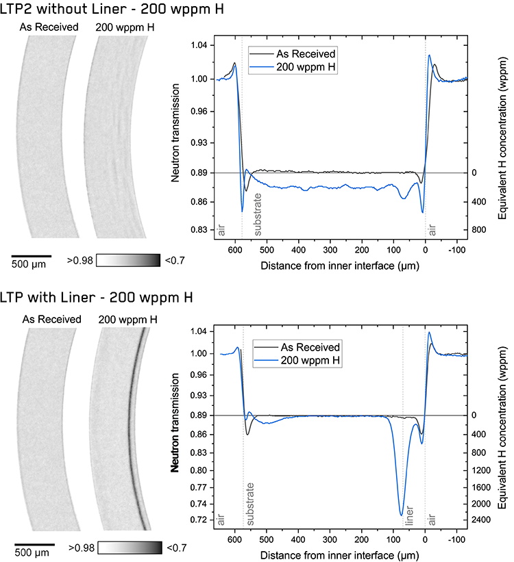 Figure 1. Neutron radiographs of the LTP2 cladding tubes without liner and the LTP cladding tubes with Fe-containing liners in the as received state and after 200 wppm of nominal hydrogen charging after homogenization with plots of their respective radial neutron transmission and corresponding hydrogen contents across the sample thickness.
Strong accumulation of hydride precipitates was observed at the interface between the liner and substrate in the LTP sample, leading to a local hydrogen concentration exceeding 2000 wppm hydrogen in the interfacial region. The transmission profiles confirm that a strong hydrogen diffusion from the bulk to the liner interface is observed for all cladding materials under different cooling rates.
At lower cooling rates, an increased fraction of the hydrides appears to nucleate and grow in the internal liner region. Samples cooled at 30 °C/h present a characteristic hydrogen-depleted zone that extends for about 200–300 µm from the liner into the substrate.
Samples quenched from the homogenization temperature of 400 °C present an increased hydrogen concentration at the liner/substrate interface. This accumulation is more pronounced in the LTP sample, where it reaches an apparent local concentration of 500 wppm. The hydrogen concentration further in the bulk of the liner of the quenched samples goes back to lower values, close to those of the substrate.
Mechanical Microscopy
Mechanical microscopy was performed over metallurgical samples over samples charged identically to those investigated by neutron radiography/microscopy. Samples both without and with liners were investigated in two conditions: as received and charged with hydrogen to 200 wppm, homogenized, and cooled at a rate of 30 °C/h were investigated. In the nanoindentation maps presented in Figure 2 the state of the four different samples is clearly observable.
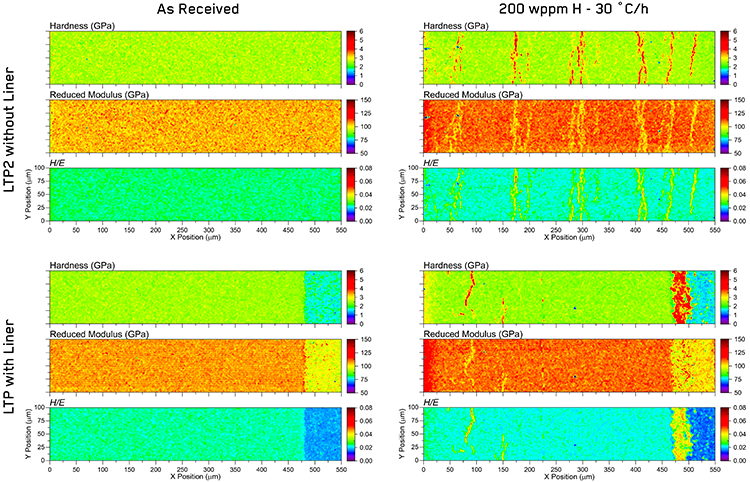 Figure 2. Nanoindentation property maps of Zr cladding tubes without and with liners in the as-received state and after an hydrogen charging of nominally 200 wppm, homogenization treatment, and followed by cooling with a rate of 30 °C/h.
The unlined, LTP2, as received sample shows mostly uniform substrate properties across the entire sample width, without edge effects noticed in the neutron radiography, with small variations corresponding to local anisotropy from individual grains. The as-received sample with a liner, LTP, shows similar properties in the substrate as the unlined sample in the majority of the cross section, but the presence of the liner is also clearly observed – which was not observable in the neutron radiographs. The liner materials are observed to be considerably softer than the substrate material used for the bulk of the cladding tube. Liner materials presented an average hardness of ~1.5 GPa; relatively soft compared to ~2.5 GPa of the substrate material. The interface between the tube and the liner is clean with no significant variations from the majority phases in either hardness or reduced modulus at the boundary.
In the samples charged with hydrogen, the presence of hydride clusters elongated and oriented in the circumferential direction is clearly visible in the indentation property maps. These present a strong contrast to the substrate properties with a ~200% increase in hardness (H = ~3.5 GPa) and a ~10% decrease in reduced modulus. The orientation and periodicity of the hydride clusters observed in the nanoindentation maps is in good agreement with the neutron radiographs. However, higher sensitivity is observed in the surface measurements made by nanoindentation than the volumetric averages quantified by neutron radiography.
In the LTP2 sample without a liner, the hydride clusters are distributed throughout the thickness of the cross-section. Whereas, in the LTP sample with a liner, the hydrides are concentrated at the liner-substrate interface, with some additional hydride clusters visible in the outer portion of the substrate beyond the hydrogen depleted zone. These outer clusters were also indicated in the neutron radiography profiles, but their interpretation was challenging due to the presence of the edge effects from neutron scattering.
As hardness and elastic modulus are interrelated properties, 2D histogram plots are utilized to display the statistical distributions of the obtained H and E values from the three different phases in the two different hydrided materials over the entire mapped regions simultaneously - Figure 3.
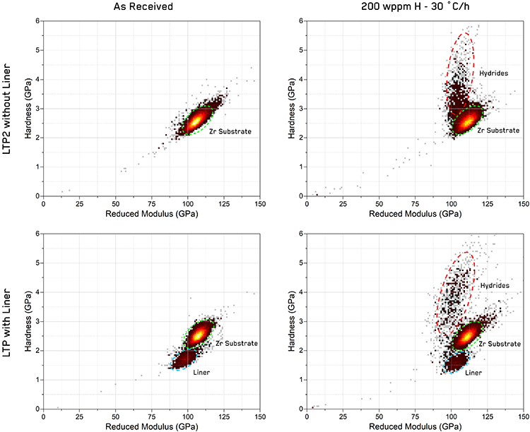 Figure 3. 2D histograms of reduced elastic modulus and hardness values obtained from nanoindentation maps presented in Figure 2 with clusters corresponding to different phases labeled.
Nanoindentation mapping allowed the identification of the hydrides as an additional phase to the substrate material and the liner material. The color of each pixel represents the number of indentations that are included within a range of H and E, which is defined as a 2D bin size. Values of only 1 indent are shown in light gray to minimize the visual impact of outliers, and higher indentation numbers are shown with a shaded gradient from darker red tones to yellow-white peaks at the highest values in each histogram. These are arbitrary units, depending on the number of total indentations performed and the bin size used.
These plots offer a simple visual method to evaluate ‘hot spots’ in the indentation property space which statistically correspond to individual phases. As shown in previous work [11], these ‘peaks’ in the 2D histograms often take the form of elliptical, normal distributions which are elongated along the direction of the H/E ratio. These can be easily segmented using mixed Gaussian clustering algorithms to measure statistical average values and standard deviations. Values for each phase extracted using cluster analysis are given in Table 1.
In all samples, the Zr substrate phase appears as a typical example of one of these clusters with a well-defined “hot spot” with values which are consistent within the standard deviation between all four samples. In the hydrogen-charged LTP2 sample, the hydride phase appears as a lobe elongated from the Zr substrate cluster with its intensity diminishing at higher hardness. This suggests that the hydrides are relatively narrow and integrated within the substrate. In the lined LTP sample, the hydride phase is more discrete from the Zr substrate with its properties more uniformly distributed producing higher average hardness values. This reflects that it occurs as a mostly separate, concentrated phase at the interface between the substrate and liner. The liner phase is also easily segmented from the majority substrate phase as a cluster with lower hardness and slightly lower modulus than the substrate phase.
Table 1. Mechanical properties extracted from cluster analysis of each sample.
|
Sample and Condition
|
Phase
|
Hardness
(GPa)
|
Reduced Modulus
(GPa)
|
Area Fraction (%)
|
|
Mean
|
SD
|
Mean
|
SD
|
--
|
|
LTP2
As Received
|
Zr Substrate
|
2.56
|
0.18
|
107.5
|
4.0
|
100
|
|
LTP2
200 wppm H – 30 °C/h
|
Zr Substrate
|
2.55
|
0.10
|
110.9
|
3.6
|
87.2
|
|
Hydride
|
3.22
|
0.74
|
105.3
|
10.3
|
12.8
|
|
LTP
As Received
|
Zr Substrate
|
2.51
|
0.16
|
107.0
|
3.2
|
84.7
|
|
Liner
|
1.70
|
0.15
|
97.6
|
4.5
|
15.3
|
|
LTP
200 wppm H – 30 °C/h
|
Zr Substrate
|
2.46
|
0.16
|
110.6
|
3.3
|
81.9
|
|
Liner
|
1.58
|
0.16
|
103.0
|
4.1
|
9.8
|
|
Hydride
|
3.80
|
0.75
|
101.9
|
7.9
|
8.3
|
Conclusion
In summary, mechanical microscopy of nuclear materials, such as hydrides at interfaces, provides valuable information about the mechanical behavior of nuclear fuel cladding alloys. Hydrides in zirconium alloys can cause embrittlement and cracking, which can lead to fuel rod failures in nuclear reactors. Therefore, understanding the distribution and mechanical properties of hydrides is important for ensuring the integrity and safety of nuclear fuel cladding materials.
Nanoindentation mapping provides local measurements of mechanical properties which allow the presence and distribution of hydrides to be characterized, as well as the effects hydrogen on the mechanical properties of the substrate and liner materials. By combining mechanical microscopy and neutron radiography, researchers can obtain a more complete picture of the relationship between hydrogen concentrations and hydride distribution and mechanical behavior. Hydrides were observed to form in clusters aligned circumferentially in the cladding tubes with preferential nucleating at the substrate-liner interface when a liner is present. Hydrides were observed to be nearly twice as hard and 10% lower in modulus than the substrate material, providing clear contrast in nanoindentation maps. This is significantly more pronounced than in volumetric measurements of the neutron radiographs, which integrate the composition of a large volume through the thickness of the cross-section, rather than the local surface measurements by nanoindentation. The hydrides at the liner interface form a distinct phase in mechanical properties, and result in a hydrogen-depleted region within the substrate. Mechanical microscopy was able to clearly resolve hydrides throughout the cross-sections, even near the edges where neutron radiography has difficulty due to scattering. Given the distinct differences in mechanical behavior, cluster algorithms were shown to easily segment the data from the various phases for statistical analysis as a function of sample conditions. This demonstrates the ability of mechanical microscopy to provide new insights into the mechanical behavior of nuclear materials such as hydrides in zirconium alloys.
Acknowledgements
The authors would like to thank Framatome for providing the materials for testing. This project has been co-financed by the Swiss Expert Group Fuels (ESB) and swissnuclear. This work is based on experiments performed at the Swiss spallation neutron source (SINQ) at Paul Scherrer Institute, Villigen, Switzerland. The authors would also like to further acknowledge and express gratitude for this collaboration within the MIDAS program.
Full details on this work are available in a publication in the Journal of Nuclear Materials: Fagnoni, L.I. Duarte, P. Trtik, J.M. Wheeler, R. Zubler & J. Bertsch. Hydrogen diffusion in zirconium cladding alloys with inner liner quantified by neutron radiography and nanoindentation, Journal of Nuclear Materials (2023).
References
- D.O. Northwood. The development and applications of zirconium alloys, Materials & Design 6 (1985) 58-70.
- C.D. Williams, M.O. Marlowe, R.A. Rand, J.S. Armijo, R.B. Adamson, S.B. Wisner. Zircaloy-2 lined zirconium barrier fuel cladding. United States: American Society for Testing and Materials, West Conshohocken, PA (United States), 1996.
- L.I. Duarte, F. Fagnoni, R. Zubler, W. Gong, P. Trtik, J. Bertsch. Effect of the inner liner on the hydrogen distribution of zircaloy-2 nuclear fuel claddings, Journal of Nuclear Materials 557 (2021) 153284.
- F. Fagnoni, P. Konarski. Hydrogen Degassing of Zirconium under High-Vacuum Conditions, Metals 12 (2022) 868.
- F. Fagnoni, L.I. Duarte, P. Trtik, J.M. Wheeler, R. Zubler, J. Bertsch. Hydrogen diffusion in zirconium cladding alloys with inner liner quantified by neutron radiography and nanoindentation, Journal of Nuclear Materials (2023).
- U. Stuhr. Time-of-flight diffraction with multiple pulse overlap. Part I: The concept, Nuclear Instruments and Methods in Physics Research Section A: Accelerators, Spectrometers, Detectors and Associated Equipment 545 (2005) 319-329.
- J. Crha, J. Vila-Comamala, E. Lehmann, C. David, P. Trtik. Light Yield Enhancement of 157-Gadolinium Oxysulfide Scintillator Screens for the High-Resolution Neutron Imaging, MethodsX 6 (2019) 107-114.
- W. Gong, P. Trtik, A.W. Colldeweih, L.I. Duarte, M. Grosse, E. Lehmann, J. Bertsch. Hydrogen diffusion and precipitation in duplex zirconium nuclear fuel cladding quantified by high-resolution neutron imaging, Journal of Nuclear Materials 526 (2019) 151757.
- W.C. Oliver, G.M. Pharr. An Improved Technique for Determining Hardness and Elastic-Modulus Using Load and Displacement Sensing Indentation Experiments, Journal of Materials Research 7 (1992) 1564-1583.
- P.S. Phani, W. Oliver. A critical assessment of the effect of indentation spacing on the measurement of hardness and modulus using instrumented indentation testing, Materials & Design 164 (2019) 107563.
- J.M. Wheeler. Mechanical phase mapping of the Taza meteorite using correlated high-speed nanoindentation and EDX, Journal of Materials Research 36 (2021) 94-104.

