Imaging on a Large Scale - Expansion Microscopy and Spatially Resolved Transcriptomics
Author: Drs. Claudia Florindo, Geraint Wilde & Mark Browne
Published: 01 Jan 2022 · Last updated: 13 Aug 2024
Recent advances in science and technology point towards the “omics” world. Omics-related sciences aim to gather information about Xn biological molecules to characterize and quantify the entire pool of molecules. Not surprisingly, microscopy is evolving to incorporate omics-related technologies, with spatially resolved transcriptomics being the latest cutting-edge microscopy approach to “Omics”.
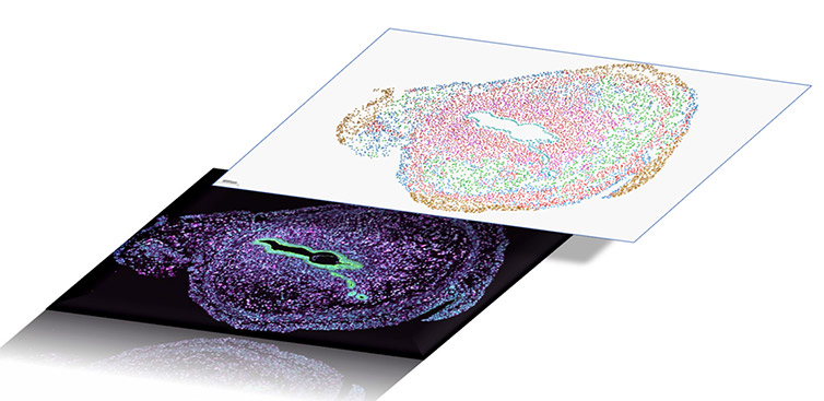
Figure 1 - Spatial Resolved Transcriptomics allows researchers to achieve an Atlas of gene expression in 2D or 3D tissues.
Concomitantly, there is an urge to gather super-resolved information to capture the detail at a single-molecule level with light microscopy imaging. Expansion Microscopy (ExM) is an imaging protocol that allows conventional light microscopes to see sub-diffraction limited (<200 nm) or densely packed details that previously could not be distinguished.
Both technologies, spatial transcriptomics and expansion microscopy need specific features in a microscope with the requirements being high-contrast imaging of large samples at high resolution and speed.
Read our poster to see why Andor Dragonfly is the ideal microscope solution for imaging spatial transcriptomics and expansion microscopy.