How to Overcome the Challenges of Imaging Vesicle Trafficking
Author: Dr Claudia Florindo
Published: 01 Aug 2019 · Last updated: 19 Dec 2022
Tags: cell-biology-lr
Challenge Background
Extracellular or intracellular vesicles perform vital functions in inter/intracellular communication. Not surprisingly, the de-regulation of the endocytic pathway affects numerous diseases, such as cancer, diabetes, cardiovascular diseases, among others. Moreover, the endocytic pathway can also be used as a valuable tool for therapeutic molecule delivery. Consequently, in depth understanding of vesicle trafficking, and the endocytic pathway is of extreme importance to promote a deeper understanding of the disease, and the development of new therapies.
Nevertheless, effectively imaging vesicle trafficking is a hard task, and researchers need to overcome several challenges to be able to do so. Key challenges to vesicle imaging are avoiding phototoxicity and photobleaching as well as the requirement for rapid acquisition speed and high imaging resolution (in either fixed cell or live imaging assays). These challenges do not end with obtaining images; post-acquisition vesicle tracking analysis is also a demanding task. Here we will focus on the speed and resolution requirements for vesicle trafficking imaging.
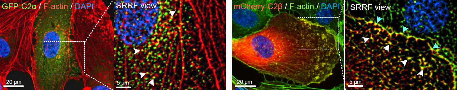
Figure 1: mCherry-C2β localizes with F-actin at the cell mebrane whereas GFP-C2α colocalizes with F-actin occassionally
Technology Solution
A camera-based confocal system is a better solution for live vesicle imaging that does not compromise cell viability due to phototoxicity. The equipment will need to acquire at fast speeds and with high imaging resolution.
Speed - The simultaneous visualization of different vesicle trafficking events requires an instrument that can acquire more than one channel at a time, and due to the fast nature of the event, the microscope will need to acquire at very fast speeds.
Resolution - The resolution required to visualize vesicle trafficking lies within the 50-100 nm window in XY. The diffraction limit of light microscopes is around 200 nm; therefore, to image vesicle trafficking, techniques that break the diffraction limit, super-resolution techniques are required.
Live cell Super-resolution - Super-resolution techniques that go beyond the diffraction limit of the light microscope generally require the acquisition of a vast number of frames (on the order of the 103 to 104 images) and /or imaging with high light intensities. In most of the cases, sample preparation is complicated, and there is also the requirement of specific fluorophores. All these requirements render most currently available Super-resolution techniques incompatible with live-cell imaging.
Andor’s Solution for Vesicle Imaging
Dragonfly is the complete solution to image intercellular trafficking, because of its speed, sensitivity and resolution. Highly sensitive cameras allow the detection of very dim signals with high Quantum Efficiency. Ultra-fast low light imaging is possible with Dragonfly and Andor´s cameras such as the back-illuminated Sona or iXon EMCCD series. Imaging modalities such as confocal spinning disk, STORM super-resolution (resolution ~ 20 nm) or TIRF (resolution ~ 50 nm) are all possible with the Dragonfly and each of these imaging modalities has advantages for vesicle research. Importantly any modality can be combined with the super-resolution technique SRRF (super-resolution radial fluctuations). SRRF-stream is compatible with live-cell imaging offering the advantage of very fast super-resolved live imaging.
Key Requirement
Vesicle imaging solution: Dragonfly and Andor´s high QE cameras
Ultra-fast acquisition speeds
The EMCCD and sCMOS detectors in the Dragonfly allow very low light imaging and high acquisition speeds. Dragonfly is at least ten times faster than point scanner confocal systems. The internal beam splitters allow Dragonfly to acquire two channels simultaneously on two independent detectors (cameras). Result 1 - Detect ultrafast events with acquisition speeds up to 400 frames per second. Result 2 - Detect two independent channels simultaneously without compromising speed or resolution.
Acquire images with a resolution of 50-100 nm
Dragonfly supports dSTORM. Dragonfly offers dual colour simultaneous TIRF with the same penetration depth. Detailed spatial information is possible. Result 1 - Resolution increase up to 20 nm with dSTORM. Result 2 - Axial resolution of 100 nm is possible with TIRF and high increase in contrast. Result 3 - Break the diffraction limit while acquiring simultaneously two independent channels.
Acquire live Super-resolved images (resolution < 200 nm)
Andor´s cameras offer an integrated licence for SRRF (Super-resolution radial fluctuations). SRRF uses the fluctuation of fluorophores emissions and their interpolation to increase the effective resolution of an optical system. Andor's built-in SRRF stream algorithm allows real-time SRRF calculations and immediate visualisation of the resolution increase. Furthermore, SRRF is compatible with conventional fluorophores, and there is no need for complex sample preparations. SRRF is compatible with confocal, TIRF and widefield imaging, being also compatible with deep imaging. Result 1 - SRRF-stream yields an increase of resolution between 2- and 6-fold (50-150 nm final resolution) in the final data. Result 2 - Due to its low power requirements (mW/cm2 to W/cm2 range), SRRF-stream is compatible with live-cell imaging. Result 3 - SRRF-stream algorithm allows acquisition of live cell images that break the diffraction limit at a frame rate of 10 frames per second.