Challenge Background
Gene manipulation for medical and biotechnological applications has been sought after since the first use of restriction enzymes in bacteria in the late 1970s. Techniques based on Zinc Finger Nucleases (ZFNs), Transcription Activator-Like Effector Nucleases (TALENs) and most notably CRISPR-Cas9, have established themselves firmly within the toolsets of many research labs.
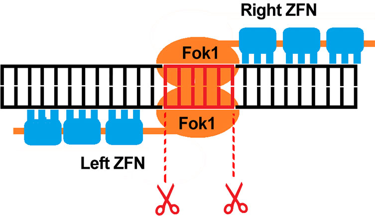
ZFNs are typically used to knockout gene functions due to insertions and deletions they can create, thereby causing disruption to coding frames
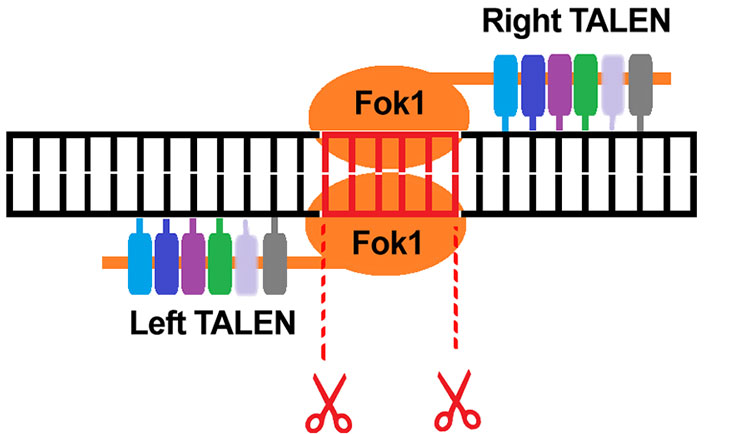
TALENS are well suited to gene editing applications such as the repair of specific genes, or insertion of gene mutations
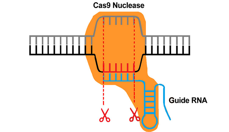
The most flexible and easy to use gene editing technique. Commonly used in gene knockout and screening applications amongst various others
CRISPR can be used to selectively target an impaired or inactive gene sequence and replace this with a functional gene. Thereby imparting the normal gene function and offering a potential route to many applications such as correcting an underlying genetic disorder in gene therapy. CRISPR based methods are now being applied beyond gene editing itself. Such studies include chromatin organization, telomere dynamics as well as epigenome editing i.e. the post translational processes associated with gene regulation. For example, silencing gene function by methylation. Highly sensitive cameras are an important part of many gene editing experiments. For example, it is challenging to visualize the fluorescent reporter tags expressed from the gene promoter sequences of successful gene constructs, or to image the organizational structure and dynamics of chromatin or of telomeres.
Technology Solution
New back-illuminated sCMOS cameras provide the exceptional sensitivity, which is required to detect the faint signals from fluorescent reporter tags like eGFP. Typically, sCMOS cameras have a large field of view and good resolution, which enables high throughput screening of multiple sgRNAs or fusion proteins in large populations of cells.
Andor Camera Solutions for Plasma Membrane Studies
Andor highly recommend the Sona back-illuminated sCMOS camera for gene editing research. The Sona 4.2B-11 offers an ideal combination of high sensitivity, exceptional field of view and speed. The high QE of 95% and low noise means that the Sona can detect the low signal levels that are inherent to these experiments. Sona 4.2B-11 provides a massive 32 mm field of view so more cells can be observed in a single image. The 11 µm pixel is perfectly suited to resolving image detail at high magnification, while use of the Andor Magnifying coupler maintains full resolution if lower magnifications such as x40 and x60 are required.
High sensitivity imaging of fluorescent reporter tags such as eGFP is important in many CRISPR based studies
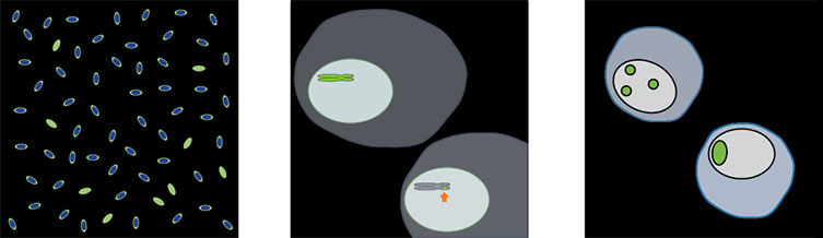
(Left to Right) Visualization of gene knockouts and validation of CRISPR gene constructs. Imaging of specific chromosomes, telomeres or genes. Live cell imaging of Chromatin structure and dynamics.
| Key Requirement |
Gene Editing Experiments Solution: Sona 4.2B-11 |
| Detect the weak signals from fluorescent reporter tags |
With 95% QE for maximum sensitivity, Sona is perfect for detecting the faint signals emitted from fluorescent reporter tags used in gene editing experiments such as drug screening. Result – Detect and confirm fluorescent tags in gene constructs. |
| Screen the largest possible number of cells |
Sona 4.2B-11 features a massive 32 mm FOV from its 4.2 Megapixel (2048x2048) sensor with 11µm pixels. This is 62% larger than competing cameras using the same sensor and over 114% larger than cameras restricted to a standard C-mount format. This means more information in each snap. Result – View the largest possible sample area and accelerate experimental throughput. |
| Resolve the spatiotemporal features of chromatin or telomeres |
The 11µm pixel of the Sona is perfectly matched for resolving the full detail of intricate chromatin spatial structure and organization as well as telomeres under high magnifications such as x100. Result – Resolve the full spatial detail at high magnification. |
| Quality and Longevity |
Sona comes with Andor’s exclusive UltraVac™ vacuum sensor enclosure. The well proven permanent vacuum process is critical not only for cooling, but for protection of the back-illuminated sensor against moisture and condensates. Result – Sustained high performance, year after year. |

