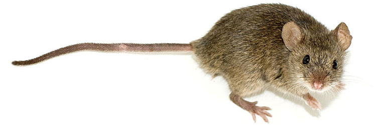
Figure 11: Image credit: George Shuklin [CC BY-SA 1.0 (https://creativecommons.org/licenses/by-sa/1.0)], from Wikimedia Commons
The mouse or M. musculus is often used as a preferred model organism due to the similarity with the human genome of 85% and genome size ~2.5 Gbp. Despite being bigger than the other model organisms discussed in this series, mice are still relatively easily to care for, breed and study.
Mice are mammals closer to our genetic makeup and have longer lifespans than the other organisms discussed in this series. As such, mice are useful for studies of more complex disorders such as ageing, neurodegenerative disorders, and modern diseases including, heart disease, obesity, high blood pressure and stress. Although issues exist due to differences in immune system behaviour between mice and humans, the mouse still proves to be the model organism of choice for many studies including cancer research [1] and neuroscience. [2]
Whether you’re using fluorescence microscopy, SIM, TIRM or two/three photon microscopy in your research, Andor has an imaging solution to meet your research needs. Andor’s portfolio of sCMOS and EMCCD detectors and software solutions could be suitable for your desired application. Please reach out to our application specialists to discuss your specific research application.
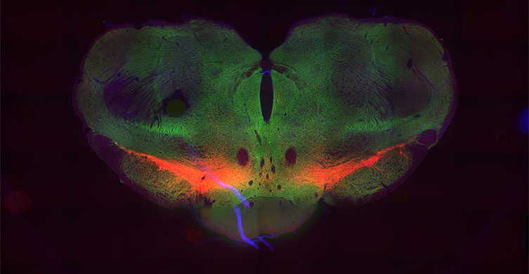
Figure 12: Mouse brain imaged with a Zyla 5.5 sCMOS camera. Image courtesy of Simon C. Watkins and Victor Tapias, Center for Biologic Imaging, University of Pittsburgh.
References
- Verdijk, T. W.J. Scheenen, W. J. Lesterhuis, G. Gambarota, A. A. Veltien, P. Walczak, N. M. Scharenborg, J. W.M. Bulte, C. J.A. Punt, A. Heerschap, C. G. Figdor, I. Jolanda and M. de Vries, Sensitivity of magnetic resonance imaging of dendritic cells for in vivo tracking of cellular cancer vaccines, Int. J. Cancer: 120, 978–984 (2006) DOI: https://doi.org/10.1002/ijc.22385
- Wang, D. G. Ouzounov, C. Wu, N. G. Horton, B. Zhang, C. Wu, Y. Zhang, M. J. Schnitzer and C. Xu, Three-photon imaging of mouse brain structure and function through the intact skull, Nature Methods, 15, 789–792 (2018), https://doi.org/10.1038/s41592-018-0115-y
Learn More from the Model Organism Series

