Advantages of using Arabidopsis Thaliana as a Model Organism
Author: Dr Alan Mullan & Dr Aleksandra Marsh
Published: 01 Feb 2019 · Last updated: 30 Jul 2025
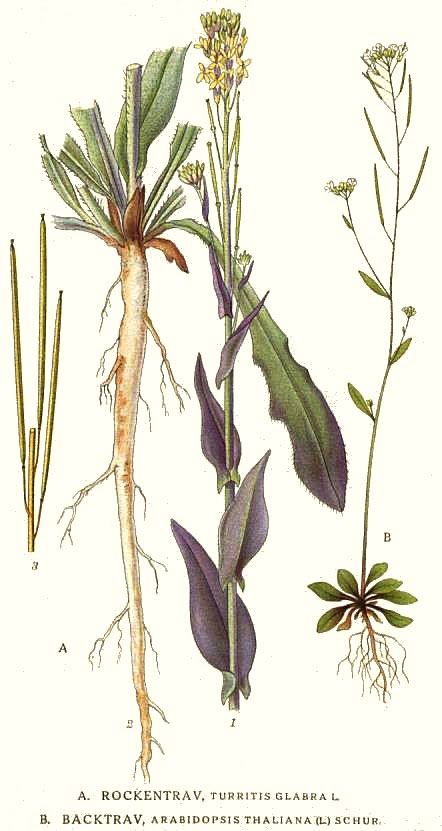
Figure 4: Painting by the Swedish botanist C. A. M. Lindman (1917-1926) A. Rockentrav, Turritis glabra L. B. Backtrav, Arabidopsis thaliana (L.) Schur.
A. thaliana, also known as rockcress or thale cress, is a small plant with white flowers, often considered to be a weed where it is found across Europe, Asia and Africa. However, in plant genetic research it is seen in a much more favourable light, being a very popular model organism for plant studies.
A.thaliana is easy to look after compared with animal model organisms. It grows quickly, produces many very small seeds, has a small genome ~114.5 Mb and is genetically well characterised due to the volume of work being focused on this plant. As a member of the Brassicaceae family it is linked to more important cultivated species such as cabbage, mustard and radish. Exposing A.thaliana to Agrobacterium tumifaciens provides a means of efficient transformation vector making A. thaliana a versatile model organism for use in the biology laboratory. A. thaliana is widely used in the fields of plant science, genetics and evolution and has helped further our understanding of germination and aspects of plant growth that are important in commercial crops.1,2 In recent years A.thaliana has even become a model organism for the study of the biochemical and molecular processes involved in human diseases.
Andor’s iKon-L camera is well suited for luminescence and modified GFP studies in reporter studies involving A.thaliana, due to its high sensitivity and low noise for experiments demanding low light conditions and long exposures.
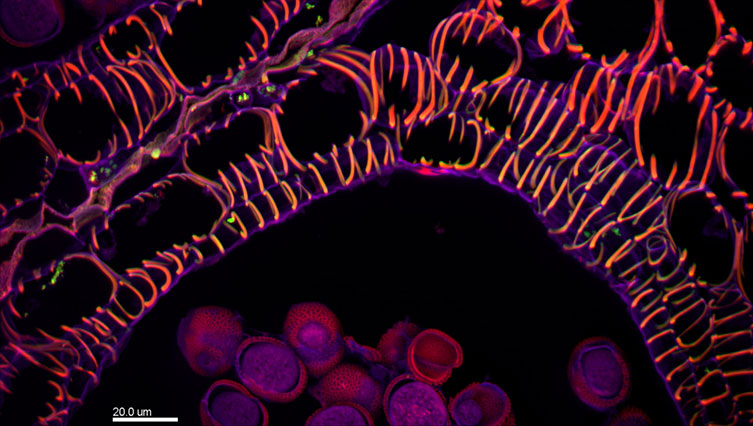
Figure 5: Autofluorescence in the flower of Brassica. Excitation at 482nm, 556nm & 640nm, Emission at 525nm (green), 609nm (red), and 676nm (blue). Maximum intensity projection from a 28um deep z-series captured at 60x magnification
References:
Endogenous Chemiluminescence from Germinating Arabidopsis Thaliana Seeds, Homa Saeidfirozeh, Azizollah Shafiekhani, Michal Cifra and Amir Ali Masoudi, Scientific Reports, (2018) 8:16231, DOI:10.1038/s41598-018-34485-6 (iXon)
Three-Dimensional Imaging of Plant Organs Using a Simple and Rapid Transparency Technique, Junko Hasegawa Yuki Sakamoto Satoru Nakagami Mitsuhiro Aida Shinichiro Sawa Sachihiro Matsunaga, Plant and Cell Physiology, Volume 57, Issue 3, 2016, Pages 462–472, https://doi.org/10.1093/pcp/pcw027 (Neo and Zyla 4.2)