September 15-16, 2021
Imaris & Dragonfly Workshop at Fred Hutchinson Cancer Research Center

Join us virtually and in-person for an Imaris and Dragonfly Workshop at Fred Hutchinson Cancer Research Center, Seattle, from Sept 15-16, 2021 where the FHCRC Cellular Imaging Shared Resource is graciously hosting the Imaris 3D/4D Visualization and Analysis teams along with the Andor Dragonfly High Speed Confocal System for live demonstrations.

We will be hosting in-person demo sessions for both, alongside an open invitation to learn from the experts and hear some great application/technical presentations on ImarisDragonfly, and more. 

In-Person Image Analysis Sessions:*

  • Advanced 3D and 4D data visualization and analysis with Imaris software, to analyze your Dragonfly acquired images.

In-Person Hands-on Dragonfly Sessions:*

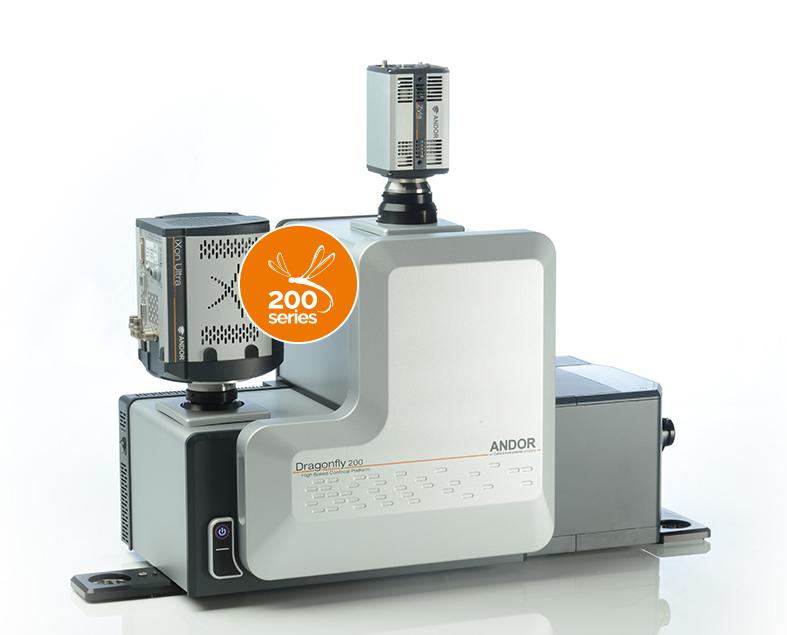
Bring your own samples or use ours and see the Dragonfly confocal system in action during an in-person demo. View the system specifications below to help prepare your sample.

Virtual Technical Presentations:*

  • Wednesday September 1, 10:00-11:00 - Andor Dragonfly - Multi-Modal High Speed Confocal Imaging
  • Wednesday September 1, 11:00-12:00 - IMARIS: Innovative Solutions for Visualizaing and Analysing 3D/4D Microscope Images

*Demo slots are planned to be in-person but are subject to change based to COVID-19 regulations. Technical presentations will be virtual unless location restrictions change. 

View the schedule and reserve your time below!

Location

Seattle, WA

Businesses

Andor, Imaris

Location & Sponsor Info

Fred Hutch Cancer Research Center
1100 Fairview Ave N, Seattle,
WA 98109, United States

Sponsor
Peng Guo, PhD
Director of Cellular Imaging Shared Resource
Fred Hutch Cancer Research Center
Cellular Imaging Core

Learn More
Contact Us

Event Schedule

Virtual Schedule

Description Location Register Date/Time
Technical Presentation
Andor Dragonfly: Multi-Modal High Speed Confocal Imaging
Online Register Here Wed Sept 1
10AM-11AM
Technical Presentation
Imaris: Innovative Solutions for Visualizing and Analysing 3D/4D Microscope Images
Online Register Here Wed Sept 1
11AM-12PM

In-Person Schedule

Description Location Register Date/Time
Imaris Image Analysis Consultation
1 to 1 sessions
In-Person Register Here Wed Sept 15
9AM - 12PM

Thurs Sept 16
9AM - 5PM
Dragonfly Demo 90min
Longer session for an in-depth experience of all functionalities
In-Person Register Here Wed Sept 15
9AM - 12PM

Thurs Sept 16
9AM - 12PM
Dragonfly Demo 1 Hour
Suited for 1 sample for an overview of the system
In-Person Register Here Thurs Sept 16
12PM - 5PM

Dragonfly based on Leica DMi8 with Mosaic and Micropoint

  • Dragonfly Imaging modalities: Spinning disk confocal, dual-camera imaging, laser widefield and deconvolution for fixed and live cells.
  • Cameras: iXon EMCCD and sCMOS Zyla
  • Lasers: 405 / 488 / 561 / 637
  • Mosaic DMD with 405nm laser: photo-activation, photo-conversion, photo-bleaching, optogenetics
  • Micropoint with 435nm laser: photostimulation, uncaging, ablation, wounding
  • Dragonfly acquires and saves images in the .ims file format, which are easily opened by Imaris 3D/4D Visualization and Analysis Software

Objectives

Immersion Mag/NA Type
Dry 10x/0.45
20x/0.75
PLAN APO
PLAN APO
Oil 63x/1.4 PLAN APO
Water 25x/0.95 (long WD)
40x/1.1
HCL FLUOTAR
PLAN APO
Glycerol 40x/1.25 PLAN APO