Nothing brings the biological electron microscopy community together like a specimen preparation problem. We have all experienced the pain of protocol optimisation; a difficult/new sample, tailoring the preparation for a specific imaging mode, or even preparing the samples in a flexible way that fits more than one microscopy modality. There are frequent discussions about this pain point in community forums and events, which I really enjoy contributing to and learning from.
For scanning electron microscopy (SEM) of biological samples, we need to ensure that the samples are stable under vacuum conditions and the electron beam, have enough contrast to visualise the structures of interest, and are conductive to avoid charging artifacts.
Stains and contrast
Biological samples are mainly composed of light elements that provide little contrast for electron microscopy. We contrast biological samples by using staining solutions with heavy elements that have high electron density. For ultrastructural imaging, elements of choice include osmium, uranium and lead. By adding these stains to biological samples, we can visualise membranes, microtubules and other large protein complexes (Fig. 1).
“There is no quality in this world that is not what it is merely by contrast.”
Herman Melville
That sounds straightforward so far, so what are the challenges I see discussed so frequently? The popular protocols can be laborious and complex, increasingly with a number of staining steps included. There is also no conformity on incubation times, temperatures, concentration of staining agents, solvents, buffers, resins… We must find ways to quantify these differences and better understand how they impact the results, with the goal of improving the efficiency of the entire process, and make results reproducible across experiments and different type of specimen.
Taking control over the methodology
Usually, the evaluation of a well prepared and stained sample is qualitative (Do I have the expected signal compared to previous experiments? Are the structures of interested distinguishable? Can I image at the desired resolution?). This means that when we are not happy with the sample preparation results, the cause of the problem might not be immediately clear. The introduction of sample characterisation using analytical methods can open the way for higher reproducibility and quantitative ways of evaluating staining.
With Energy Dispersive Spectroscopy (EDS), it is possible to measure and map stains and native elements in biological samples, in both TEM and SEM, in an easy, fast, and non-destructive manner (introduced in the standard imaging process). With quantitative data for studying contrast generation, there is room for streamlining specimen preparation techniques. EDS unlocks the possibility to address questions such as:
- How is sample contrast affected by staining concentration?
- Do different cells and tissues have the same affinity for stains?
- What is my subcellular stain distribution? Is there selective staining?
- Is my protocol reproducible?
Without extra experimental burden, EDS opens the way for better quality control and additional ways to interpret electron microscopy of cells and tissues.
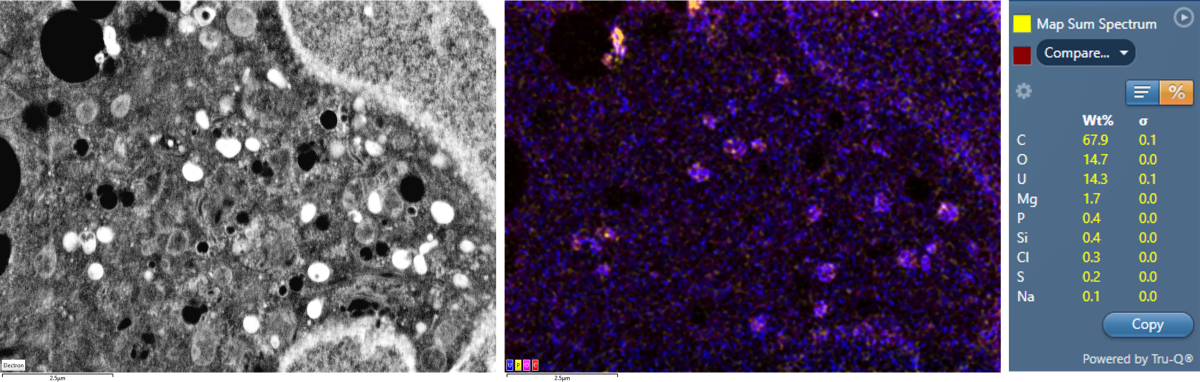
Figure 1 – Cell stained with uranyl acetate. EDS maps shows overlayed uranium (U) colocalised with phosphate (P) and carboxyl (C, O) groups. Quantification table expressed in weight percentage (wt%).
Beyond SEM
Most biological complex questions require the use of more than a single imaging technique. With multimodal approaches, it is crucial to consider a flexible specimen preparation which does not compromise any of the selected imaging methods.
To learn more about how and why to choose the preparation methods that are compatible with multiple imaging workflows, please register for the webinar and watch it live or on demand.


